Review
. 2019 May 3:7:73.
doi: 10.3389/fcell.2019.00073. eCollection 2019.

Supramolecular Complexes in Cell Death and Inflammation and Their Regulation by Autophagy

Affiliations
Review

Supramolecular Complexes in Cell Death and Inflammation and Their Regulation by Autophagy

Ian E Gentle. Front Cell Dev Biol. .

Abstract

Signaling activation is a tightly regulated process involving myriad posttranslational modifications such as phosphorylation/dephosphorylation, ubiquitylation/deubiquitylation, proteolytical cleavage events as well as translocation of proteins to new compartments within the cell. In addition to each of these events potentially regulating individual proteins, the assembly of very large supramolecular complexes has emerged as a common theme in signal transduction and is now known to regulate many signaling events. This is particularly evident in pathways regulating both inflammation and cell death/survival. Regulation of the assembly and silencing of these complexes plays important roles in immune signaling and inflammation and the fate of cells to either die or survive. Here we will give a summary of some of the better studied supramolecular complexes involved in inflammation and cell death, particularly with a focus on diseases caused by their autoactivation and the role autophagy either plays or may be playing in their regulation.

Keywords: RHIM; autophagy; cargo receptors; cell death; death domain; inflammation; innate immunity; supramolecular complexes.


Figures
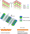
FIGURE 1
FIGURE 1
Structural elements of death domain family, RHIM and ubiquitin scaffolds. (A) Death domain family interaction can form fibrillar structures. Death domain family members interact with themselves through three different interaction modes known as Types I–III, depending on the surfaces of the domain that interact. Shown are two possible helical fibrillar assemblies using differing combinations of interfaces. Multiple forms of this type of interaction have been identified, allowing for modular assembly of different complexes. (B) RHIM–RHIM scaffolds form amyloid fibrils. Shown is a fibril of two proteins containing a RHIM and globular domains. The fibril is formed by two parallel beta amyloid sheets coming together. This brings the globular domains into close proximity for interaction and potential activation such as kinase domains of RIPK1/3. RHIM fibrils can be mixed or homogeneous (RIPK1–RIPK3 or RIPK3–RIPK3 fibers form example). (C) Polyubiquitin chains have different functional roles. Shown are K48 and K63/linear ubiquitin chains. The structural layout of the individual chains is different resulting in recruitment of different ubiquitin binding proteins. K63 and linear ubiquitin chains are similar in their layout, although still functionally distinct. K48 chains are predominantly used for proteasomal degradation, whereas K63 and linear ubiquitin chains are used for recruitment of NF-κB activating complexes such as TAB/TAK and IKK as well as linking to autophagic cargo receptors among other functions.
FIGURE 2
FIGURE 2
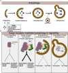
Autophagy regulates turnover of Supramolecular complexes. (A) Summary of general autophagic process. The autophagic machinery including LC3 is recruited to the donour membrane and forms the phagophore. This then extends to form the isolation membrane which begins to engulf cytoplasmic or ubiquitylated contents. Eventually the enclosed autophagosome is formed which then can fuse with lysosomes to form the autolysosome. Lysosomal enzymes then degrade the contents of the autolysosome. (B) Summary of specific autophagy mediated by cargo receptors. Assembled supramolecular signaling complexes begin their signaling response. Cargo receptors such as p62 are recruited via K63 ubiquitin chains on the complex. TBK1 which has been activated and recruited to the complex phosphorylates the cargo receptor to enhance its recruitment. At this stage the signal from the complex may be amplified due to clustering of multiple complexes together or perhaps further enhancement of the localized proximity of kinases, caspases, and ubiquitin ligases for example. As the cargo:cargo-receptor complexes are engulfed by the forming autophagosome, signaling will be reduced. Finally, degradation of the complex within the autolysosome completes the cycle and the complex is destroyed.
FIGURE 3
FIGURE 3
Supramolecular signaling complexes in inflammation share common scaffolds and signaling pathways. The supramolecular signaling complexes formed by TLR, TNFSR, NOD1/2, STING, MAVS, Carma, BCL-10, MALT1 (CBM) and inflammasome complexes are indicated. Shown are the core scaffolds formed through death domain family interactions as well as RHIM interactions. STING contains no death domain family member or RHIM. Specific proteins known to interact through death domain interactions and RHIM interaction are indicated within each complex. Recruitment of TBK1 and/or K63/linear ubiquitin is indicated. TBK1 recruitment activates IRF3 to induce interferon responses. K63/linear ubiquitin recruits the TAB/TAK and IKK complexes which result in activation of NF-kB and Map kinase transcriptional responses. Also shown is linear/K63 ubiquitination of ASC of inflammasomes which can promote their assembly and activation or degradation. Complexes and organelles such as mitochondria that are k63 ubiquitylated recruit autophagy cargo receptors such as p62. This leads ultimately to degradation and silencing of the complexes. This is enhanced by TBK1 mediated phosphorylation of the cargo receptors. Other scaffolds such as those mediated via TRAF proteins are also present, but for simplicity have been omitted from the figure. DD family and RHIM scaffold are not meant to be to scale or reflect the actual organization of the scaffold, but simply indicate that each of these scaffolds are present.
FIGURE 4
FIGURE 4
Supramolecular signaling complexes can activate cell death. (Upper) The indicated complexes and the respective scaffolds recruited are indicated. Numbers correspond to the modes of cell death shown in the lower panel ad indicate the modes shown to be triggered by the respective complexes. (Lower) Different modes of cell death and the role of the respective scaffold in activating caspases, or RIP kinases is shown. (1) Inflammasomes assemble a core filament of ASC which in turn recruits filaments of caspase-1, to cluster it and trigger its activation. Active caspase-1 then cleaves Gasdermin D to allow the cleaved form to assemble into multimeric pores in the plasma membrane, causing pyroptosis. (2) Release of cytochrome C through Bax/Bak pores on the mitochondrial outer membrane triggers formation of the apoptosome. The apoptosome consists of a core of APAF1 and caspase-9 interacting through CARD–CARD interactions as indicated. This allows activation of caspase-9. Caspase-9 then cleaves and activates caspase-3/7 to trigger apoptosis. (3) Caspase-8 clustering onto FADD causes its autoactivation. Clustering is mediated through DED–DED interactions between FADD and caspase-8 and caspase-8 itself (not shown is cFLIP which can commingle with caspase-8 to regulate its activation). Active caspase-8 can cleave caspase-3/7 and trigger apoptosis. This scaffold can be activated by numerous receptors as indicated. (4) Recruitment and activation of RIPK1/3 through RHIM–RHIM mediated amyloid fibers can trigger FADD recruitment and clustering through the DD of RIPK1. Clustered FADD recruits caspase-8, probably causing similar aggregation as shown in mode 3. Active caspase-8 cleaves caspase-3/7 to trigger apoptosis. (5) The kinase domains of RIPK1 and RIPK3 are also brought into close proximity by RHIM fibers. Normally, the capase-8 indicated in mode 4 will inactivate these through cleavage of their kinase domains, however, when no caspase-8 activity is present, as is shown, RIPK3 is activated and phosphorylates MLKL. Phosphorylated MLKL assembles into pore complexes in the plasma membrane, causing necroptosis.

Similar articles

Cited by

References

    1. Abe T., Barber G. N. (2014). Cytosolic-DNA-mediated, STING-dependent proinflammatory gene induction necessitates canonical NF-κB activation through TBK1. J. Virol. 88 5328–5341. 10.1128/JVI.00037-14 - DOI - PMC - PubMed
    1. Ablasser A., Goldeck M., Cavlar T., Deimling T., Witte G., Röhl I., et al. (2013). cGAS produces a 2′-5′-linked cyclic dinucleotide second messenger that activates STING. Nature 498 380–384. 10.1038/nature12306 - DOI - PMC - PubMed
    1. Aliprantis A. O., Yang R. B., Weiss D. S., Godowski P., Zychlinsky A. (2000). The apoptotic signaling pathway activated by Toll-like receptor-2. EMBO J. 19 3325–3336. 10.1093/emboj/19.13.3325 - DOI - PMC - PubMed
    1. Anand P. K., Tait S. W. G., Lamkanfi M., Amer A. O., Nunez G., Pages G., et al. (2011). TLR2 and RIP2 pathways mediate autophagy of Listeria monocytogenes via extracellular signal-regulated kinase (ERK) activation. J. Biol. Chem. 286 42981–42991. 10.1074/jbc.M111.310599 - DOI - PMC - PubMed
    1. Ando M., Kawazu M., Ueno T., Fukumura K., Yamato A., Soda M., et al. (2013). Cancer-associated missense mutations of caspase-8 activate nuclear factor-κB signaling. Cancer Sci. 104 1002–1008. 10.1111/cas.12191 - DOI - PMC - PubMed

LinkOut - more resources