Review
. 2019 May 3:6:31.
doi: 10.3389/fmolb.2019.00031. eCollection 2019.

Melting Down Protein Stability: PAPS Synthase 2 in Patients and in a Cellular Environment

Affiliations
Review

Melting Down Protein Stability: PAPS Synthase 2 in Patients and in a Cellular Environment

Oliver Brylski et al. Front Mol Biosci. .

Abstract

Within the crowded and complex environment of the cell, a protein experiences stabilizing excluded-volume effects and destabilizing quinary interactions with other proteins. Which of these prevail, needs to be determined on a case-by-case basis. PAPS synthases are dimeric and bifunctional enzymes, providing activated sulfate in the form of 3'-phosphoadenosine-5'-phosphosulfate (PAPS) for sulfation reactions. The human PAPS synthases PAPSS1 and PAPSS2 differ significantly in their protein stability as PAPSS2 is a naturally fragile protein. PAPS synthases bind a series of nucleotide ligands and some of them markedly stabilize these proteins. PAPS synthases are of biomedical relevance as destabilizing point mutations give rise to several pathologies. Genetic defects in PAPSS2 have been linked to bone and cartilage malformations as well as a steroid sulfation defect. All this makes PAPS synthases ideal to study protein unfolding, ligand binding, and the stabilizing and destabilizing factors in their cellular environment. This review provides an overview on current concepts of protein folding and stability and links this with our current understanding of the different disease mechanisms of PAPSS2-related pathologies with perspectives for future research and application.

Keywords: PAPS synthase; enzyme storage complex; excluded volume effect; ligand stabilization; quinary interaction; sulfation pathways.


Figures
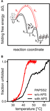
Figure 1
Figure 1
A theoretical and an experimental perspective on protein folding. (A) Schematic representation of the folding free energy landscape for a multi-state (black) and two-state folder (red). Kinetic constants for folding and unfolding (kf and ku) are depicted with arrows highlighting the transitions between unfolded (U), intermediate (I), and folded (F) states. (B) Thermal CD unfolding data of PAPS synthase 2 adapted from van den Boom et al. (2012). Experiments without (red) and with (black) the APS nucleotide at a 100 fold excess of APS. Transitions observed fit the expected data for a two-state folder with one transition and a three-state folder with two distinguishable transitions. Multi-state behavior is only observed upon APS binding.
Figure 2
Figure 2
The catalytic cycle of APS kinase. Schematic representation including substrate binding and product release steps. An inhibitory or stable storage complex is populated after PAPS release by binding of APS (lower left).
Figure 3
Figure 3
Structural mapping of PAPSS2 point mutations. Known disease-relevant mutations in PAPS synthase 2 isoform (red spheres) are shown in a PAPS synthase structure (PDB: 1XNJ). The N-terminal APS kinase is shown in gray, the C-terminal ATP-sulfurylase domain in black. Zoom, disease relevant mutations in the APS kinase central beta-sheet (black). Amino acid numbering reflects positions in PAPSS2. Table, solvent accessible surface area (SASA) of sidechains derived from DSSP values. Values for amino acids in the APS kinase domain have been derived from PDB structure 2AX4, values for the ATP sulfurylase are from 1XNJ.

Similar articles

Cited by

References

    1. Alberti S., Gladfelter A., Mittag T. (2019). Considerations and challenges in studying liquid-liquid phase separation and biomolecular condensates. Cell 176, 419–434. 10.1016/j.cell.2018.12.035 - DOI - PMC - PubMed
    1. Alfano C., Sanfelice D., Martin S. R., Pastore A., Temussi P. A. (2017). An optimized strategy to measure protein stability highlights differences between cold and hot unfolded states. Nat. Commun. 8:15428. 10.1038/ncomms15428 - DOI - PMC - PubMed
    1. Arosio P., Vendruscolo M., Dobson C. M., Knowles T. P. (2014). Chemical kinetics for drug discovery to combat protein aggregation diseases. Trends Pharmacol. Sci. 35, 127–135. 10.1016/j.tips.2013.12.005 - DOI - PubMed
    1. Ataullakhanov F. I., Vitvitsky V. M. (2002). What determines the intracellular ATP concentration. Biosci. Rep. 22, 501–511. 10.1023/A:1022069718709 - DOI - PubMed
    1. Batra J., Xu K., Qin S., Zhou H. X. (2009). Effect of macromolecular crowding on protein binding stability: modest stabilization and significant biological consequences. Biophys. J. 97, 906–911. 10.1016/j.bpj.2009.05.032 - DOI - PMC - PubMed