

Lower Blood Pressure Is Not Associated With Decreased Arterial Spin Labeling Estimates of Perfusion in Intracerebral Hemorrhage
- PMID: 31131671
- PMCID: PMC6585347
- DOI: 10.1161/JAHA.118.010904
Lower Blood Pressure Is Not Associated With Decreased Arterial Spin Labeling Estimates of Perfusion in Intracerebral Hemorrhage
Abstract
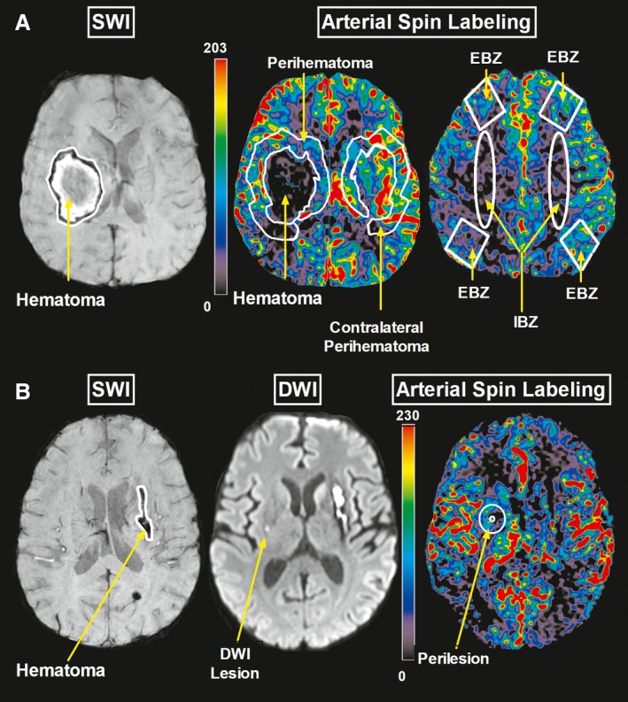
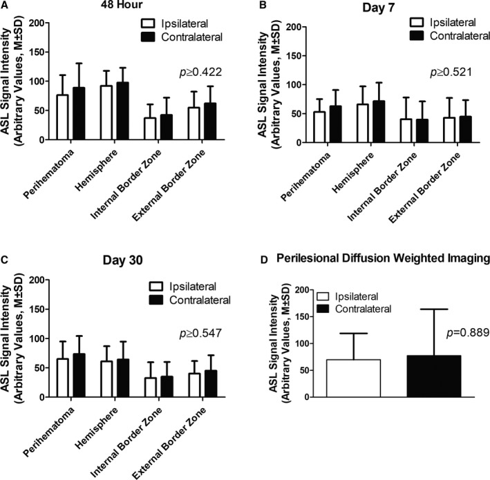
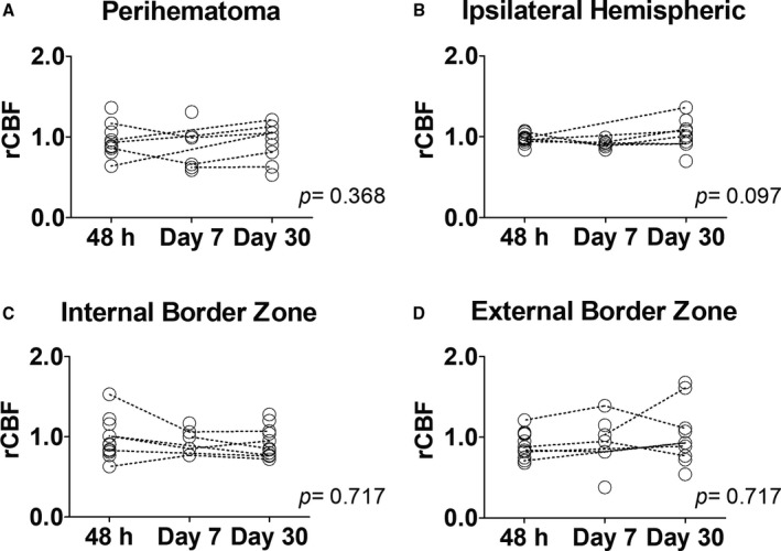
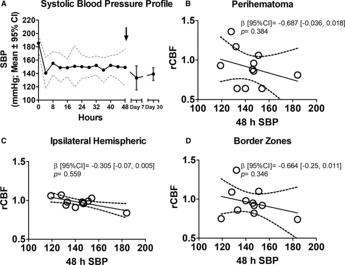
Background Subacute ischemic lesions in intracerebral hemorrhage ( ICH ) have been hypothesized to result from hypoperfusion. Although studies of cerebral blood flow ( CBF ) indicate modest hypoperfusion in ICH , these investigations have been limited to early time points. Arterial spin labeling ( ASL ), a magnetic resonance imaging technique, can be used to measure CBF without a contrast agent. We assessed CBF in patients with ICH using ASL and tested the hypothesis that CBF is related to systolic blood pressure ( SBP ). Methods and Results In this cross-sectional study, patients with ICH were assessed with ASL at 48 hours, 7 days, and/or 30 days after onset. Relative CBF ( rCBF ; ratio of ipsilateral/contralateral perfusion) was measured in the perihematomal regions, hemispheres, border zones, and the perilesional area in patients with diffusion-weighted imaging hyperintensities. Twenty-patients (65% men; mean± SD age, 68.5±12.7 years) underwent imaging with ASL at 48 hours (N=12), day 7 (N=6), and day 30 (N=11). Median (interquartile range) hematoma volume was 13.1 (6.3-19.3) mL. Mean± SD baseline SBP was 185.4±25.5 mm Hg. Mean perihematomal rCBF was 0.9±0.2 at 48 hours at all time points. Baseline SBP and other SBP measurements were not associated with a decrease in rCBF in any of the regions of interest ( P≥0.111). r CBF did not differ among time points in any of the regions of interest ( P≥0.097). Mean perilesional rCBF was 1.04±0.65 and was unrelated to baseline SBP ( P=0.105). Conclusions ASL can be used to measure rCBF in patients with acute and subacute ICH . Perihematomal CBF was not associated with SBP changes at any time point. Clinical Trial Registration URL: http://www.clinicaltrials.gov . Unique identifier: NCT00963976.
Keywords: arterial spin labeling; blood pressure; intracerebral hemorrhage; magnetic resonance imaging.
Figures
Similar articles
-
Cerebral perfusion and blood pressure do not affect perihematoma edema growth in acute intracerebral hemorrhage.Stroke. 2014 May;45(5):1292-8. doi: 10.1161/STROKEAHA.113.003194. Epub 2014 Apr 1. Stroke. 2014. PMID: 24692481 Clinical Trial.
-
The Intracerebral Hemorrhage Acutely Decreasing Arterial Pressure Trial.Stroke. 2013 Mar;44(3):620-6. doi: 10.1161/STROKEAHA.111.000188. Epub 2013 Feb 7. Stroke. 2013. PMID: 23391776 Clinical Trial.
-
Acute blood pressure reduction in patients with intracerebral hemorrhage does not result in borderzone region hypoperfusion.Stroke. 2014 Oct;45(10):2894-9. doi: 10.1161/STROKEAHA.114.005614. Epub 2014 Aug 21. Stroke. 2014. PMID: 25147326 Clinical Trial.
-
Perfusion imaging by arterial spin labeling in migraine: A literature review.J Cereb Blood Flow Metab. 2024 Aug;44(8):1253-1270. doi: 10.1177/0271678X241237733. Epub 2024 Mar 14. J Cereb Blood Flow Metab. 2024. PMID: 38483125 Free PMC article. Review.
-
Changes in Cerebral Blood Flow and Diffusion-Weighted Imaging Lesions After Intracerebral Hemorrhage.Transl Stroke Res. 2022 Oct;13(5):686-706. doi: 10.1007/s12975-022-00998-6. Epub 2022 Mar 19. Transl Stroke Res. 2022. PMID: 35305264 Review.
References
-
- Anderson CS, Heeley E, Huang Y, Wang J, Stapf C, Delcourt C, Lindley R, Robinson T, Lavados P, Neal B, Hata J, Arima H, Parsons M, Li Y, Wang J, Heritier S, Li Q, Woodward M, Simes RJ, Davis SM, Chalmers J. Rapid blood‐pressure lowering in patients with acute intracerebral hemorrhage. N Engl J Med. 2013;368:2355–2365. - PubMed
-
- Hanley DF, Hsu CY, Martin RL, Moy CS, Silbergleit R, Steiner T, Suarez JI, Toyoda K, Wang Y, Yamamoto H, Yoon B‐WW, Qureshi AI, Palesch YY, Barsan WG, Hanley DF, Hsu CY, Martin RL, Moy CS, Silbergleit R, Steiner T, Suarez JI, Toyoda K, Wang Y, Yamamoto H, Yoon B‐WW; ATACH‐2 Trial Investigators and the Neurological Emergency Treatment Trials Network . Intensive blood‐pressure lowering in patients with acute cerebral hemorrhage. N Engl J Med. 2016;375:1033–1043. - PMC - PubMed
-
- Butcher KS, Baird T, MacGregor L, Desmond P, Tress B, Davis S. Perihematomal edema in primary intracerebral hemorrhage is plasma derived. Stroke. 2004;35:1879–1885. - PubMed
Publication types
MeSH terms
Substances
Associated data
LinkOut - more resources
Full Text Sources
Medical
