

Lineage tracing suggests that ovarian endosalpingiosis does not result from escape of oviductal epithelium
- PMID: 31131879
- PMCID: PMC7457448
- DOI: 10.1002/path.5308
Lineage tracing suggests that ovarian endosalpingiosis does not result from escape of oviductal epithelium
Abstract
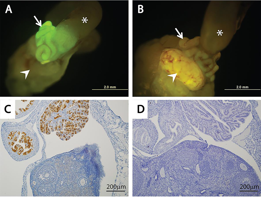
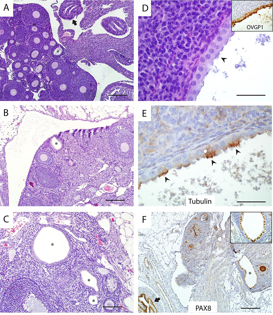
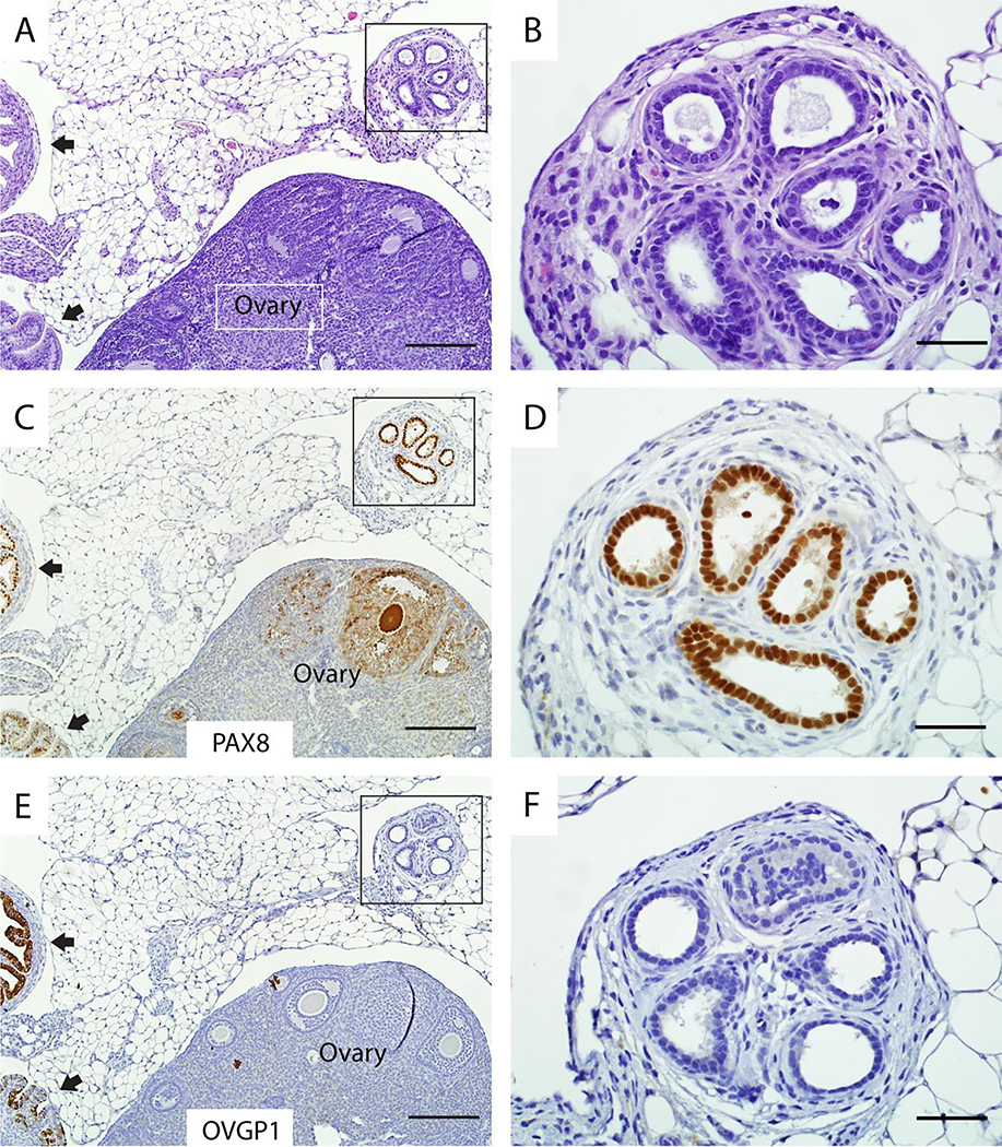
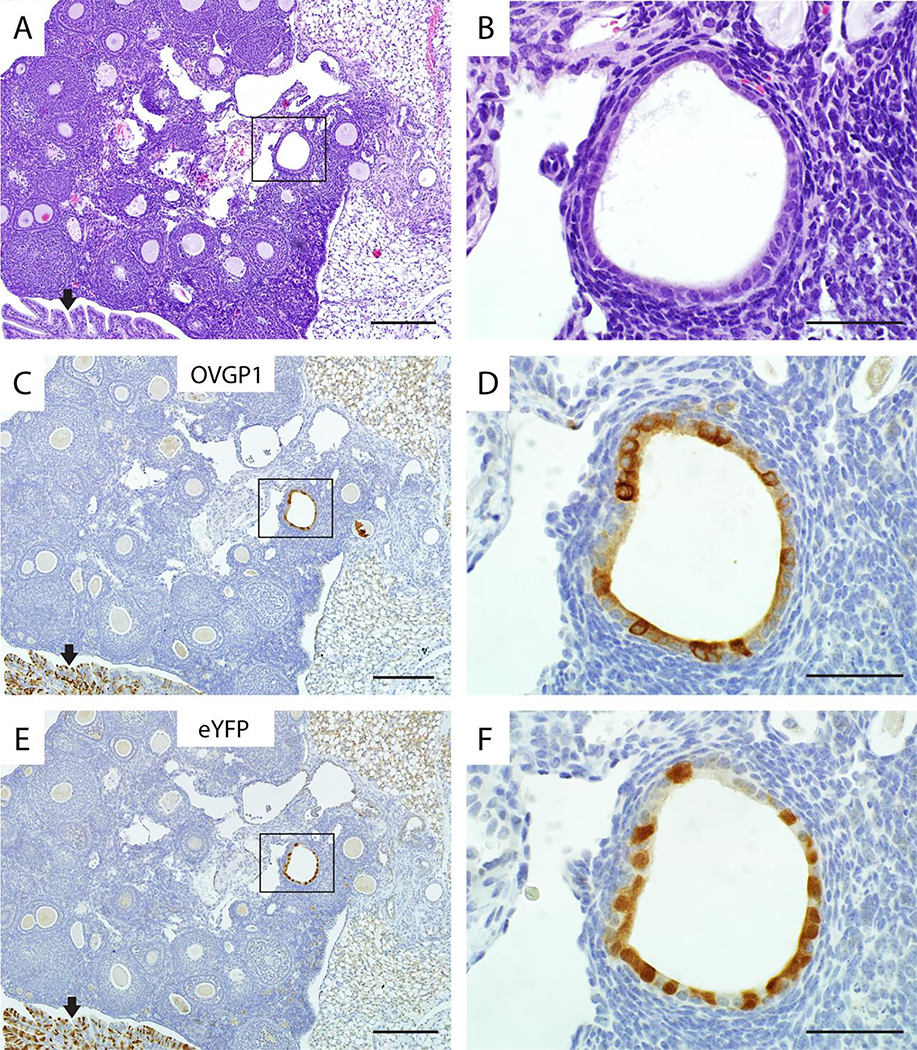
Most high-grade serous carcinomas are thought to arise from Fallopian tube epithelium (FTE), but some likely arise outside of the tube, perhaps from ectopic tubal-type epithelium known as endosalpingiosis. Importantly, the origin of endosalpingiosis is poorly understood. The proximity of the tubal fimbriae to the ovaries has led to the proposal that disruptions in the ovarian surface that occur during ovulation may allow detached FTE to implant in the ovary and form tubal-type glands and cysts. An alternative model suggests that cells present in ectopic locations outside the Müllerian tract retain the capacity for multi-lineage differentiation and can form glands with tubal-type epithelium. We used double transgenic Ovgp1-iCreERT2 ;R26RLSL-eYFP mice, which express an eYFP reporter protein in OVGP1-positive tissues following transient tamoxifen (TAM) treatment, to track the fate of oviductal epithelial cells. Cohorts of adult mice were given TAM to activate eYFP expression in oviductal epithelium, and ovaries were examined at time points ranging from 2 days to 12 months post-TAM. To test whether superovulation might increase acquisition of endosalpingiosis, additional cohorts of TAM-treated mice underwent up to five cycles of superovulation and ovaries were examined at 1, 6, and 12 months post-TAM. Ovaries were sectioned in their entirety to identify endosalpingiosis. Immunohistochemical staining for PAX8, tubulin, OVGP1, and eYFP was employed to study endosalpingiosis lesions. Ovarian endosalpingiosis was identified in 14.2% of TAM-treated adult mice. The endosalpingiotic inclusion glands and cysts were lined by secretory and ciliated cells and expressed PAX8, tubulin, OVGP1, and eYFP. Neither age nor superovulation was associated with a significant increase in endosalpingiosis. Endosalpingiosis was also occasionally present in the ovaries of pre-pubertal mice. The findings imply that ovarian endosalpingiosis in the mouse does not likely arise as a consequence of detachment and implantation of tubal epithelium and other mechanisms may be relevant. © 2019 Pathological Society of Great Britain and Ireland. Published by John Wiley & Sons, Ltd.
Keywords: Fallopian tube; endosalpingiosis; high-grade serous carcinoma; lineage tracing; mouse model.
© 2019 Pathological Society of Great Britain and Ireland. Published by John Wiley & Sons, Ltd.
Conflict of interest statement
Figures
References
-
- Piek JM, van Diest PJ, Zweemer RP, et al. Dysplastic changes in prophylactically removed Fallopian tubes of women predisposed to developing ovarian cancer. J Pathol 2001; 195: 451–456. - PubMed
-
- Crum CP, Drapkin R, Miron A, et al. The distal fallopian tube: a new model for pelvic serous carcinogenesis. Curr Opin Obstet Gynecol 2007; 19: 3–9. - PubMed
-
- Kindelberger DW, Lee Y, Miron A, et al. Intraepithelial carcinoma of the fimbria and pelvic serous carcinoma: Evidence for a causal relationship. Am J Surg Pathol 2007; 31: 161–169. - PubMed
-
- Lee Y, Miron A, Drapkin R, et al. A candidate precursor to serous carcinoma that originates in the distal fallopian tube. J Pathol 2007; 211: 26–35. - PubMed
-
- Medeiros F, Muto MG, Lee Y, et al. The tubal fimbria is a preferred site for early adenocarcinoma in women with familial ovarian cancer syndrome. Am J Surg Pathol 2006; 30: 230–236. - PubMed
Publication types
MeSH terms
Substances
Grants and funding
- P30 CA046592/CA/NCI NIH HHS/United States
- P30CA046592/National Cancer Institute at the National Institutes of Health/International
- R01 CA196619/CA/NCI NIH HHS/United States
- K07 CA096619/CA/NCI NIH HHS/United States
- R01CA196619/Transgenic Animal Models and Biostatistics, Analytics & Bioinformatics Shared Resources/International
LinkOut - more resources
Full Text Sources
Medical
Molecular Biology Databases
Miscellaneous
