

Nucleotide-binding oligomerization domain-like receptor X1 restricts porcine reproductive and respiratory syndrome virus-2 replication by interacting with viral Nsp9
- PMID: 31132368
- PMCID: PMC7114581
- DOI: 10.1016/j.virusres.2019.05.011
Nucleotide-binding oligomerization domain-like receptor X1 restricts porcine reproductive and respiratory syndrome virus-2 replication by interacting with viral Nsp9
Abstract
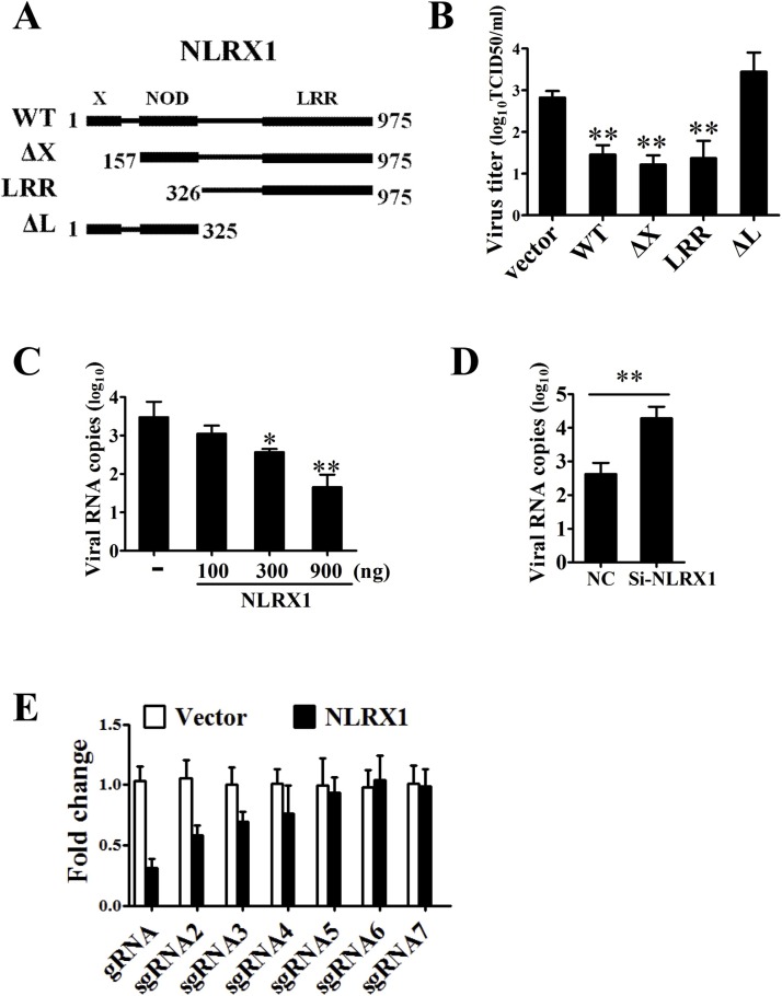
Porcine reproductive and respiratory syndrome virus (PRRSV) causes one of the most economically important diseases of swine worldwide. Current antiviral strategies provide only limited protection. Nucleotide-binding oligomerization domain-like receptor (NLR) X1 is unique among NLR proteins in its functions as a pro-viral or antiviral factor to different viral infections. To date, the impact of NLRX1 on PRRSV infection remains unclear. In this study, we found that PRRSV infection promoted the expression of NLRX1 gene. In turn, ectopic expression of NLRX1 inhibited PRRSV replication in Marc-145 cells, whereas knockdown of NLRX1 enhanced PRRSV propagation in porcine alveolar macrophages (PAMs). Mechanistically, NLRX1 was revealed to impair intracellular viral subgenomic RNAs accumulation. Finally, Mutagenic analyses indicated that the LRR (leucine-rich repeats) domain of NLRX1 interacted with PRRSV Nonstructural Protein 9 (Nsp9) RdRp (RNA-dependent RNA Polymerase) domain and was necessary for antiviral activity. Thus, our study establishes the role of NLRX1 as a new host restriction factor in PRRSV infection.
Keywords: Nonstructural Protein 9 (Nsp9); Nucleotide-binding oligomerization domain-like receptor X1 (NLRX1); PRRSV-host interactions; Porcine reproductive and respiratory syndrome virus (PRRSV); Replication.
Copyright © 2019 Elsevier B.V. All rights reserved.
Figures


Similar articles
-
Interleukin-2 enhancer binding factor 2 interacts with the nsp9 or nsp2 of porcine reproductive and respiratory syndrome virus and exerts negatively regulatory effect on the viral replication.Virol J. 2017 Jul 11;14(1):125. doi: 10.1186/s12985-017-0794-5. Virol J. 2017. PMID: 28693575 Free PMC article.
-
ZAP, a CCCH-Type Zinc Finger Protein, Inhibits Porcine Reproductive and Respiratory Syndrome Virus Replication and Interacts with Viral Nsp9.J Virol. 2019 May 1;93(10):e00001-19. doi: 10.1128/JVI.00001-19. Print 2019 May 15. J Virol. 2019. PMID: 30867303 Free PMC article.
-
The DEAD-box RNA helicase 5 positively regulates the replication of porcine reproductive and respiratory syndrome virus by interacting with viral Nsp9 in vitro.Virus Res. 2015 Jan 2;195:217-24. doi: 10.1016/j.virusres.2014.10.021. Epub 2014 Nov 1. Virus Res. 2015. PMID: 25449571 Free PMC article.
-
Mechanisms of suppression of interferon production by porcine reproductive and respiratory syndrome virus.Acta Virol. 2012;56(1):3-9. doi: 10.4149/av_2012_01_3. Acta Virol. 2012. PMID: 22404603 Review.
-
The PRRSV replicase: exploring the multifunctionality of an intriguing set of nonstructural proteins.Virus Res. 2010 Dec;154(1-2):61-76. doi: 10.1016/j.virusres.2010.07.030. Epub 2010 Aug 7. Virus Res. 2010. PMID: 20696193 Free PMC article. Review.
Cited by
-
Research progress on the pattern recognition receptors involved in porcine reproductive and respiratory syndrome virus infection.Front Cell Infect Microbiol. 2024 Aug 15;14:1428447. doi: 10.3389/fcimb.2024.1428447. eCollection 2024. Front Cell Infect Microbiol. 2024. PMID: 39211800 Free PMC article. Review.
-
NLRX1 Mediates the Disruption of Intestinal Mucosal Function Caused by Porcine Astrovirus Infection via the Extracellular Regulated Protein Kinases/Myosin Light-Chain Kinase (ERK/MLCK) Pathway.Cells. 2024 May 25;13(11):913. doi: 10.3390/cells13110913. Cells. 2024. PMID: 38891045 Free PMC article.
-
Screening of Porcine Innate Immune Adaptor Signaling Revealed Several Anti-PRRSV Signaling Pathways.Vaccines (Basel). 2021 Oct 14;9(10):1176. doi: 10.3390/vaccines9101176. Vaccines (Basel). 2021. PMID: 34696285 Free PMC article.
-
ZNF283, a Krüppel-associated box zinc finger protein, inhibits RNA synthesis of porcine reproductive and respiratory syndrome virus by interacting with Nsp9 and Nsp10.Vet Res. 2024 Jan 15;55(1):9. doi: 10.1186/s13567-023-01263-w. Vet Res. 2024. PMID: 38225617 Free PMC article.
-
NLRX1 Is a Multifaceted and Enigmatic Regulator of Immune System Function.Front Immunol. 2019 Oct 11;10:2419. doi: 10.3389/fimmu.2019.02419. eCollection 2019. Front Immunol. 2019. PMID: 31681307 Free PMC article. Review.
References
-
- Abdul-Sater A.A., Said-Sadier N., Lam V.M., Singh B., Pettengill M.A., Soares F., Tattoli I., Lipinski S., Girardin S.E., Rosenstiel P., Ojcius D.M. Enhancement of reactive oxygen species production and chlamydial infection by the mitochondrial Nod-like family member NLRX1. J. Biol. Chem. 2010;285(53):41637–41645. - PMC - PubMed
-
- Allen I.C., Moore C.B., Schneider M., Lei Y., Davis B.K., Scull M.A., Gris D., Roney K.E., Zimmermann A.G., Bowzard J.B., Ranjan P., Monroe K.M., Pickles R.J., Sambhara S., Ting J.P. NLRX1 protein attenuates inflammatory responses to infection by interfering with the RIG-I-MAVS and TRAF6-NF-kappaB signaling pathways. Immunity. 2011;34(6):854–865. - PMC - PubMed
-
- Cao S.F., Guo Q.Y., Wang Y. Inhibition of highly pathogenic porcine reproductive and respiratory syndrome virus replication by recombinant pseudorabies virus-mediated RNA interference in piglets. Vet. Microbiol. 2015;181(3-4):212–220. - PubMed
Publication types
MeSH terms
Substances
LinkOut - more resources
Full Text Sources
Research Materials
