Review
. 2019 Aug:103:305-315.
doi: 10.1016/j.neubiorev.2019.05.016. Epub 2019 May 24.

Establishing a framework for neuropathological correlates and glymphatic system functioning in Parkinson's disease

Affiliations
Review

Establishing a framework for neuropathological correlates and glymphatic system functioning in Parkinson's disease

Saranya Sundaram et al. Neurosci Biobehav Rev. 2019 Aug.

Abstract

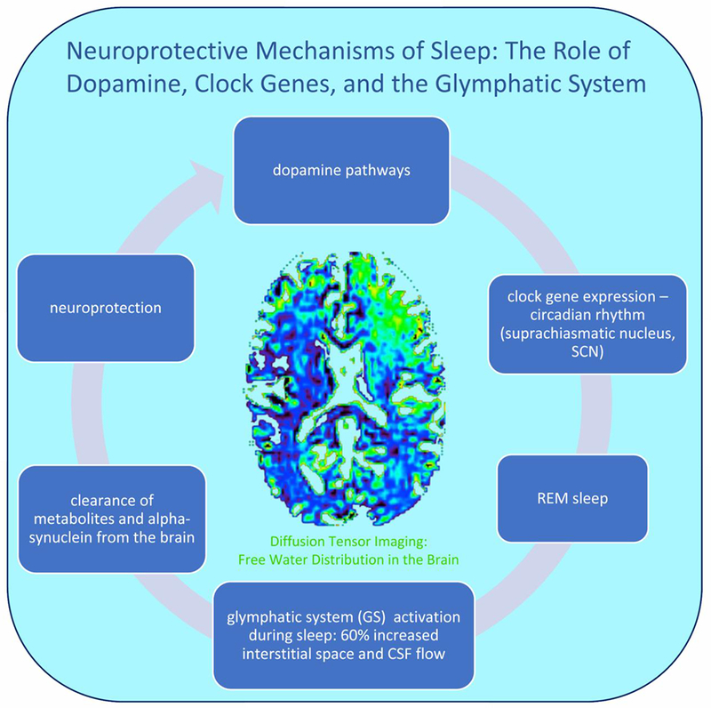
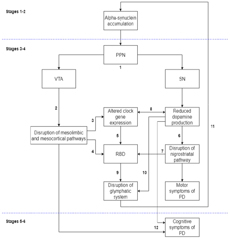
Recent evidence has advanced our understanding of the function of sleep to include removal of neurotoxic protein aggregates via the glymphatic system. However, most research on the glymphatic system utilizes animal models, and the function of waste clearance processes in humans remains unclear. Understanding glymphatic function offers new insight into the development of neurodegenerative diseases that result from toxic protein inclusions, particularly those characterized by neuropathological sleep dysfunction, like Parkinson's disease (PD). In PD, we propose that glymphatic flow may be compromised due to the combined neurotoxic effects of alpha-synuclein protein aggregates and deteriorated dopaminergic neurons that are linked to altered REM sleep, circadian rhythms, and clock gene dysfunction. This review highlights the importance of understanding the functional role of glymphatic system disturbance in neurodegenerative disorders and the subsequent clinical and neuropathological effects on disease progression. Future research initiatives utilizing noninvasive brain imaging methods in human subjects with PD are warranted, as in vivo identification of functional biomarkers in glymphatic system functioning may improve clinical diagnosis and treatment of PD.

Keywords: Alpha-synuclein; Clock genes; Dopamine; Glymphatic system; Parkinson’s disease; REM-sleep behavior disorder.


Conflict of interest statement

Declarations of Interests

None.

Figures

Figure 1.
Figure 1.. Neuroprotective mechanisms of sleep.
Midbrain dopamine neurons innervate corticolimbic and nigrostriatal systems involved in the control of sleep and wake states, including the SCN (circadian rhythm) (Monti and Monti, 2007) and striatal clock gene expression (Verwey et al., 2016) that promote healthy sleep. During sleep, glymphatic system activity is characterized by a 60% increase in the interstitial space and CSF flow (Jessen et al., 2015; Xie et al., 2013), and clearance of alpha-synuclein, along with other protein accumulations, from the brain (Iliff et al., 2012). Non-invasive free-water diffusion tensor imaging (FW-DTI) can be utilized in human subjects to detect the percent of free water in the tissue during sleep and wake states (Thomas et al., 2018).
Figure 2.
Figure 2.. Proposed model of prodromal factors in the development of Parkinson’s disease.
A pathological model for understanding the neurodegenerative progression of PD has been proposed by Braak and colleagues (2003, 2004) based on post-mortem analysis of brain tissue samples. Briefly, stages 1 and 2 of their model are characterized by the early development of alpha-synuclein rich Lewy bodies within lower brainstem regions and the olfactory bulb and represent a “preclinical” phase of the disease. Stages 3 and 4 mark the onset of clinical symptoms in PD due to spreading pathology to several key mid- and forebrain regions largely implicated in PD. including the substantia nigra (SN) and pedunculopontine nucleus (PPN), among others. Stages 5 and 6 are thought to represent the end stages of disease progression as Lewy bodies spread diffusely up through the cortex. We thus propose that the clinical and other biological manifestations of PD occur within the context of the Braak staging model. (1) As noted, alpha-synuclein accrual in prodromal PD occurs in dopaminergic-rich brainstem regions including the SN and PPN, the latter of which projects to both the ventral tegmental area (VTA) and the SN (Braak et al., 2003; French and Muthusamy, 2018). (2) Deterioration of the VTA leads to the disruption of mesolimbic and mesocortical dopaminergic pathways (Le Moal and Simon, 1991; Westerink and Kwint, 1996), which in turn (3) alter circadian functioning (McClung, 2007) and (4) promote disturbances in sleep through REM-sleep behavior disorder (RBD; Chaudhuri and Schapira, 2009; Fantini and Ferini-Strambi, 2007; Kalaitzakis et al., 2013; Rye, 2004). (5) Altered clock gene expression is highly implicated in the development of several forms of sleep disturbances, including RBD (Turek et al., 2001). (6) Concurrent deterioration of the SN leads to disruption of the nigrostriatal pathway via dopaminergic depletion, and (7) is implicated in RBD (Kim et al., 2010). (8) Altered clock gene expression decreases dopamine production (Albrecht, 2013), and reduced dopamine levels in turn blunt daily rhythms in clock gene expression (Cai et al., 2010). (9) We propose that disrupted sleep via RBD that is perpetuated by dysregulated clock gene expression, (10) along with dopaminergic dysfunction that promote effective waste clearance (Gratwicke et al., 2015; Jennings and Rusakov, 2016), reduces opportunities for glymphatic system functioning and (11) contributes to alpha-synuclein buildup in perivascular pathways, thereby exacerbating neurodegeneration and emerging disease symptoms (Musiek and Holtzman, 2016). Continued mechanistic disturbances in dopaminergic pathways, sleep, and glymphatic clearance of alpha-synuclein over time perpetuate the course of PD development, (12) leading to later clinical signs of PD, such as cognitive decline.

References

    1. Aarsland Bronnick K, Williams-Gray C, Weintraub D, Marder K, Kulisevsky J, Bum D, Barone P, Pagonabarraga J, Allcock L, Santangelo G, Foltynie T, Janvin C, Larsen JP, Barker RA, Emre M, 2010. Mild cognitive impairment in Parkinson disease: A multicenter pooled analysis. Neurology 75, 1062–1069. 10.1212/WNL.0b013e3181f39d0e - DOI - PMC - PubMed
    1. Agnati LF, Genedani S, Lenzi PL, Leo G, Mora F, Ferre S, Fuxe K, 2005. Energy gradients for the homeostatic control of brain ECF composition and for VT signal migration: Introduction of the tide hypothesis. J. Neural Transm. 112, 45–63. 10.1007/s00702-004-0180-5 - DOI - PubMed
    1. Ahlskog JE, 2007. Beating a dead horse: Dopamine and Parkinson disease. Neurology 69, 1701–1711. 10.1212/01.wnl.0000296942.14309.4a - DOI - PubMed
    1. Alberico SL, Cassell MD, Narayanan NS, 2015. The vulnerable ventral tegmental area in Parkinson’s disease. Basal Ganglia 5, 51–55. 10.1016/j.baga.2015.06.001 - DOI - PMC - PubMed
    1. Albin RL, Young AB, Penney JB, 1989. The functional anatomy of basal ganglia disorders. Trends Neurosci. 12, 366–375. 10.1016/0166-2236(89)90074-X - DOI - PubMed

Publication types

MeSH terms