

Review
doi: 10.1016/j.sbi.2019.03.028.
Epub 2019 May 25.
Development of photolabile protecting groups and their application to the optochemical control of cell signaling
Affiliations
- PMID: 31132552
- PMCID: PMC7026702
- DOI: 10.1016/j.sbi.2019.03.028
Item in Clipboard
Review
Development of photolabile protecting groups and their application to the optochemical control of cell signaling
Curr Opin Struct Biol.
2019 Aug.
Abstract
Many biological processes are naturally regulated with spatiotemporal control. In order to perturb and investigate them, optochemical tools have been developed that convey similar spatiotemporal precision. Pivotal to optochemical probes are photolabile protecting groups, so called caging groups, and recent developments have enabled new applications to cellular processes, including cell signaling. This review focuses on the advances made in the field of caging groups and their application in cell signaling through caged molecules such as neurotransmitters, lipids, secondary messengers, and proteins.
Copyright © 2019. Published by Elsevier Ltd.
Figures
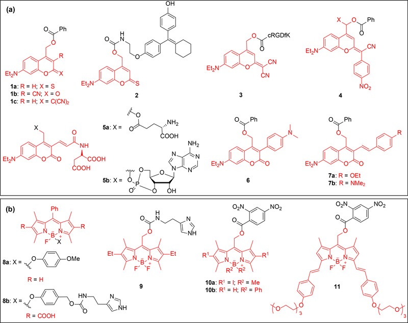
Structures of coumarin and BODIPY caged substrates; caging groups are shown in red. (a) Structures include D-π-A type (1-5) and D-π-D (6-7) coumarin caged substrates. The structures include caged benzoic acids (1, 4, 6–7), a caged tamoxifen analogue (2), a caged cyclic RGDfK peptide (3), caged glutamic acid (5a) and caged cAMP (5b). (b) BODIPY caged molecules include 4-methoxyphenol (8a), caged histamine (8b, 9), and caged 2,4-dinitrobenzoic acid (10-11).
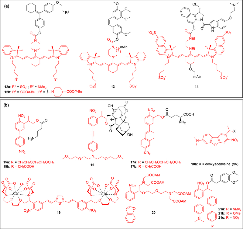
Structures of heptamethine cyanine and new nitrobenzyl (NB) caged substrates; caging groups are shown in red. (a) Caged substrates include 4-hydroxycycofen (12), combretastatin A4 (13), and duocarmycin (14), the last two substrates were conjugated to the antibody panitumumab. (b) Structures include NPE-type (17-18 and 21) and NPP-type (15-16) caged molecules. Caged substrates include GABA (15), gibberellic acid (16), glutamic acid (17), deoxyadenosine (18), calcium chelators (19-20), and 3,4-dimethoxyphenylacetic acid (21);
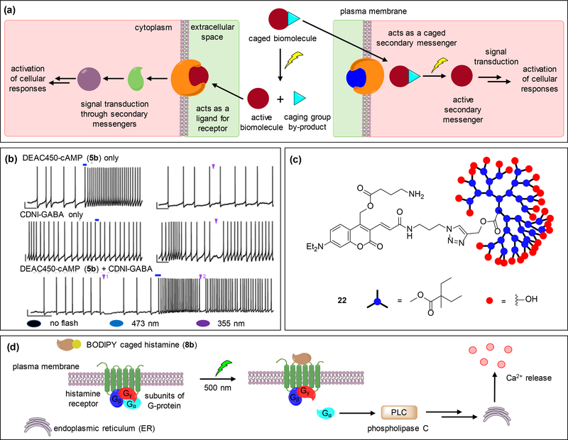
Examples of applications of coumarin and BODIPY caging groups in cell-signaling. (a) The general approach includes a caged inactive biomolecule that is rendered functional upon light irradiation. The active biomolecule can act as a receptor ligand or a secondary messenger, both of which induce downstream signaling events and lead to a cellular response. (b) Whole cell current clamp recordings demonstrate the wavelength-selective, bi-directional modulation of neuronal firing in striatal cholinergic neurons through decaging of DEAC450-cAMP (5b) and CDNI-GABA within the same cell using one-photon irradiation. While decaging of 5b causes increase in the firing rates, decaging of CDNI-GABA transiently inhibits it. (c) Dendrimer-cloaked DEAC450-GABA (22) shields the substrate from interacting with the GABA-A receptors thereby reducing background activity. (d) Upon irradiation of BODIPY-caged histamine 8b, histamine binds to the H1 receptor and induces the G-protein mediated activation of phospholipase C, leading to calcium release. Adapted with permission from ref. [31], Copyright 2013 American Chemical Society.
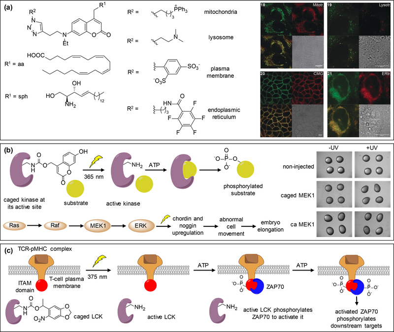
Use of nitrobenzyl and coumarin caging groups in the control of cell signaling. (a) Organelle targeting DEACM-caged arachidonic acid and sphingosine derivatives are localized as expected: mitochondria (upper left), lysosome (upper right), plasma membrane (lower left), and endoplasmic reticulum (lower right). (b) A kinase is rendered inactive through caging of a catalytic site lysine with 7-hydroxycoumarin, blocking ATP binding until exposure to 365 nm irradiation. The active kinase then phosphorylates its downstream substrate. This was applied to MEK1 within the Ras/MAPK signaling pathway in developing zebrafish embryos. Hyperactivation of MEK1 induces embryo dorsalization leading to an elongation phenotype at 8 hours post fertilization. (c) LCK is rendered inactive through caging of its catalytic site until light irradiation with 375 nm activates the enzyme, which in turn activates ITAMs through phosphorylation. ZAP70 gets recruited by ITAMs and phosphorylated by LCK to activate downstream effector molecules and induce multiple signaling pathways. Adapted with permission from ref. [72] and [73], Copyright 2018, John Wiley and Sons and Copyright 2017, American Chemical Society, respectively.
References
Publication types
MeSH terms
Substances
Grants and funding
LinkOut - more resources
Full Text Sources
