Review
. 2019 Jun;20(6):863-879.
doi: 10.3348/kjr.2018.0450.

Emerging Role of Hepatobiliary Magnetic Resonance Contrast Media and Contrast-Enhanced Ultrasound for Noninvasive Diagnosis of Hepatocellular Carcinoma: Emphasis on Recent Updates in Major Guidelines

Affiliations
Review

Emerging Role of Hepatobiliary Magnetic Resonance Contrast Media and Contrast-Enhanced Ultrasound for Noninvasive Diagnosis of Hepatocellular Carcinoma: Emphasis on Recent Updates in Major Guidelines

Tae Hyung Kim et al. Korean J Radiol. 2019 Jun.

Abstract

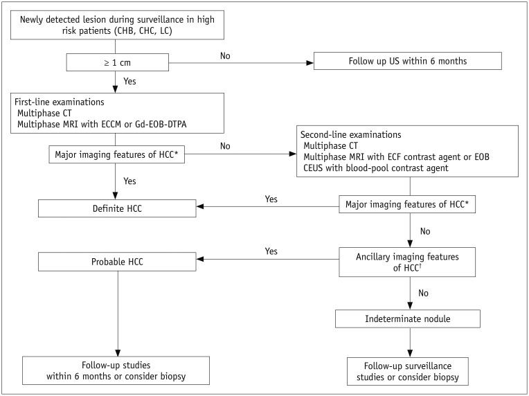
Hepatocellular carcinoma (HCC) can be noninvasively diagnosed on the basis of its characteristic imaging findings of arterial phase enhancement and portal/delayed "washout" on computed tomography (CT) and magnetic resonance imaging (MRI) in cirrhotic patients. However, different specific diagnostic criteria have been proposed by several countries and major academic societies. In 2018, major guideline updates were proposed by the Association for the Study of Liver Diseases, European Association for the Study of the Liver (EASL), Korean Liver Cancer Association and National Cancer Center (KLCA-NCC) of Korea. In addition to dynamic CT and MRI using extracellular contrast media, these new guidelines now include magnetic resonance imaging (MRI) using hepatobiliary contrast media as the first-line diagnostic test, while the KLCA-NCC and EASL guidelines also include contrast-enhanced ultrasound (CEUS) as the second-line diagnostic test. Therefore, hepatobiliary MR contrast media and CEUS will be increasingly used for the noninvasive diagnosis and staging of HCC. In this review, we discuss the emerging role of hepatobiliary phase MRI and CEUS for the diagnosis of HCC and also review the changes in the HCC diagnostic criteria in major guidelines, including the KLCA-NCC practice guidelines version 2018. In addition, we aimed to pay particular attention to some remaining issues in the noninvasive diagnosis of HCC.

Keywords: Criteria; Diagnosis; Hepatocellular carcinoma; Imaging techniques; Practice guidelines.


Conflict of interest statement

The authors have no potential conflicts of interest to disclose.

Figures

Fig. 1
Fig. 1. Typical CEUS features of HCC in 60-year-old man with history of right hemihepatectomy for HCC.
On arterial (A) and delayed (B) phases of CT, 2.3-cm subtle low-attenuated nodule (arrow) is visible only in delayed phase at left lateral segment of liver. Nodule (arrow) shows low echogenicity on gray-scale US (C). On CEUS, nodule (arrowheads) shows hyperenhancement in arterial phase (25 seconds after contrast injection) (D), isoechogenicity in portal phase (45 seconds) (E), and mild washout occurring only in late phase (200 seconds) (F), compatible with LI-RADS 5. CEUS = contrast-enhanced ultrasound, CT = computed tomography, HCC = hepatocellular carcinoma, LI-RADS = Liver Imaging Reporting and Data System, US = ultrasound
Fig. 2
Fig. 2. Typical CEUS findings of cholangiocarcinoma in 57-year-old woman with CHB viral infection.
On arterial (A) and portal (B) phases of CT, 8-cm low-attenuated mass (arrows) is seen with equivocal hyperenhancement in arterial phase and central hyperenhancement with peripheral low attenuation in 3-minute delayed phase at right lobe of liver. Mass (arrowheads) shows isoechogenicity on gray-scale US (C). On CEUS, mass (arrowheads) shows heterogeneous central hyperenhancement in arterial phase (20 seconds after contrast injection) (D), marked washout in both portal (60 seconds) (E) and late (180 seconds) (F) phases, compatible with LI-RADS-M. Lesion was confirmed as cholangiocarcinoma via biopsy. CHB = chronic hepatitis B
Fig. 3
Fig. 3. Diagnostic algorithm for suspected HCC using new KLCA-NCC practice guidelines.
*Major imaging features of HCC include arterial hyperenhancement and washout appearance during portal venous, delayed, or HBP on multiphasic CT or MRI using extracellular contrast agents or EOB in nodules ≥ 1 cm in diameter. However, lesion should not show either marked T2 high SI or targetoid appearance on DWI or contrast-enhanced sequences. On CEUS as second-line examinations, major imaging features include arterial hyperenhancement and late (≥ 60 seconds) and mild washout, In nodule(s) with some but not all aforementioned major imaging features of HCC, category of “probable” HCC can be assigned only when lesion fulfills at least one item from each of following two categories of ancillary imaging features. Two categories which make up ancillary imaging features are findings favoring malignancy in general (mild-to-moderate T2 hyperintensity, restricted diffusion, HBP hypointensity, interval growth) and those favoring HCC in particular (non-enhancing capsule, mosaic architecture, nodule-in-nodule appearance, fat or blood products in mass). These criteria should be applied only to lesion which shows neither marked T2 hyperintensity nor targetoid appearance on DWI or contrast-enhanced sequences. DWI = diffusion-weighted imaging, ECCM = extracellular contrast media, Gd-EOB-DTPA = gadolinium ethoxybenzyl diethylenetriamine pentaacetic acid, HBP = hepatobiliary phase, KLCA-NCC = Korean Liver Cancer Association-National Cancer Center, LC = liver cirrhosis, MRI = magnetic resonance imaging, SI = signal intensity
Fig. 4
Fig. 4. Gadoxetic acid-enhanced MRI in 62-year-old man with CHB.
On arterial phase MRI (A), 1.2-cm enhancing nodule is visualized in S4 (arrows) abutting middle hepatic vein. Nodule shows iso-, iso-, and hypointensity on portal (B); transitional (C); and hepatobiliary (D) phases, respectively. Lesion was pathologically confirmed as HCC. This nodule does not meet criteria of LR-5 according to Western guidelines; however, this nodule can be diagnosed as HCC according to updated 2018 KLCA-NCC guidelines.
Fig. 5
Fig. 5. Gadoxetic acid-enhanced MRI in 70-year-old man with CHB.
On T2-weighted (A) and precontrast T1-weighted (B) MRI, 2-cm nodule (arrow) is seen with marked T2 hyperintensity and low T1 SI at subcapsular portion of segment 2 of liver. After contrast injection, lesion demonstrates nodular enhancement in arterial phase (C) and persistent enhancement in portal phase (D). Lesion (arrowheads) depicts hypointensity on HBP (E). Lesion was proved to be hemangioma by showing no interval change over years. Regardless of arterial enhancement with hepatobiliary defect, diagnosis of HCC cannot be made due to exclusion criteria of marked T2 hyperintensity according to updated KLCA-NCC guidelines version 2018.
Fig. 6
Fig. 6. Gadoxetic acid-enhanced MRI in 70-year-old woman with chronic hepatitis C.
On fat-saturated T2-weighted image (A), approximately 5-cm mass (arrow) with mild hyperintensity is seen in segment 4 of liver. After gadoxetic acid injection, lesion (arrows) shows arterial hyperenhancement (B), isointensity on portal phase (C), and hypointensity on HBP (D). On diffusion-weighted image (E), mass (arrowheads) demonstrates peripheral hyperintensity with typical targetoid appearance. Mass was histopathologically confirmed as cholangiocarcinoma. Regardless of presence of arterial enhancement with hepatobiliary defect, diagnosis of HCC cannot be made due to exclusion criteria of targetoid appearance according to updated KLCA-NCC guidelines version 2018.

Similar articles

Cited by

References

    1. Choo SP, Tan WL, Goh BKP, Tai WM, Zhu AX. Comparison of hepatocellular carcinoma in Eastern versus Western populations. Cancer. 2016;122:3430–3446. - PubMed
    1. Altekruse SF, McGlynn KA, Reichman ME. Hepatocellular carcinoma incidence, mortality, and survival trends in the United States from 1975 to 2005. J Clin Oncol. 2009;27:1485–1491. - PMC - PubMed
    1. Singal AG, Pillai A, Tiro J. Early detection, curative treatment, and survival rates for hepatocellular carcinoma surveillance in patients with cirrhosis: a meta-analysis. PLoS Med. 2014;11:e1001624. - PMC - PubMed
    1. Trevisani F, De Notariis S, Rapaccini G, Farinati F, Benvegnù L, Zoli M, et al. Semiannual and annual surveillance of cirrhotic patients for hepatocellular carcinoma: effects on cancer stage and patient survival (Italian experience) Am J Gastroenterol. 2002;97:734–744. - PubMed
    1. Ando E, Kuromatsu R, Tanaka M, Takada A, Fukushima N, Sumie S, et al. Surveillance program for early detection of hepatocellular carcinoma in Japan: results of specialized department of liver disease. J Clin Gastroenterol. 2006;40:942–948. - PubMed