. 2019 May 27;17(1):54.
doi: 10.1186/s12964-019-0358-y.

Crystal structure of a cytocidal protein from lamprey and its mechanism of action in the selective killing of cancer cells

Affiliations

Crystal structure of a cytocidal protein from lamprey and its mechanism of action in the selective killing of cancer cells

Yue Pang et al. Cell Commun Signal. .

Abstract

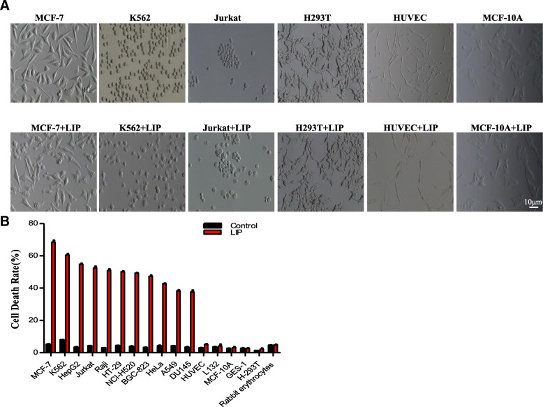
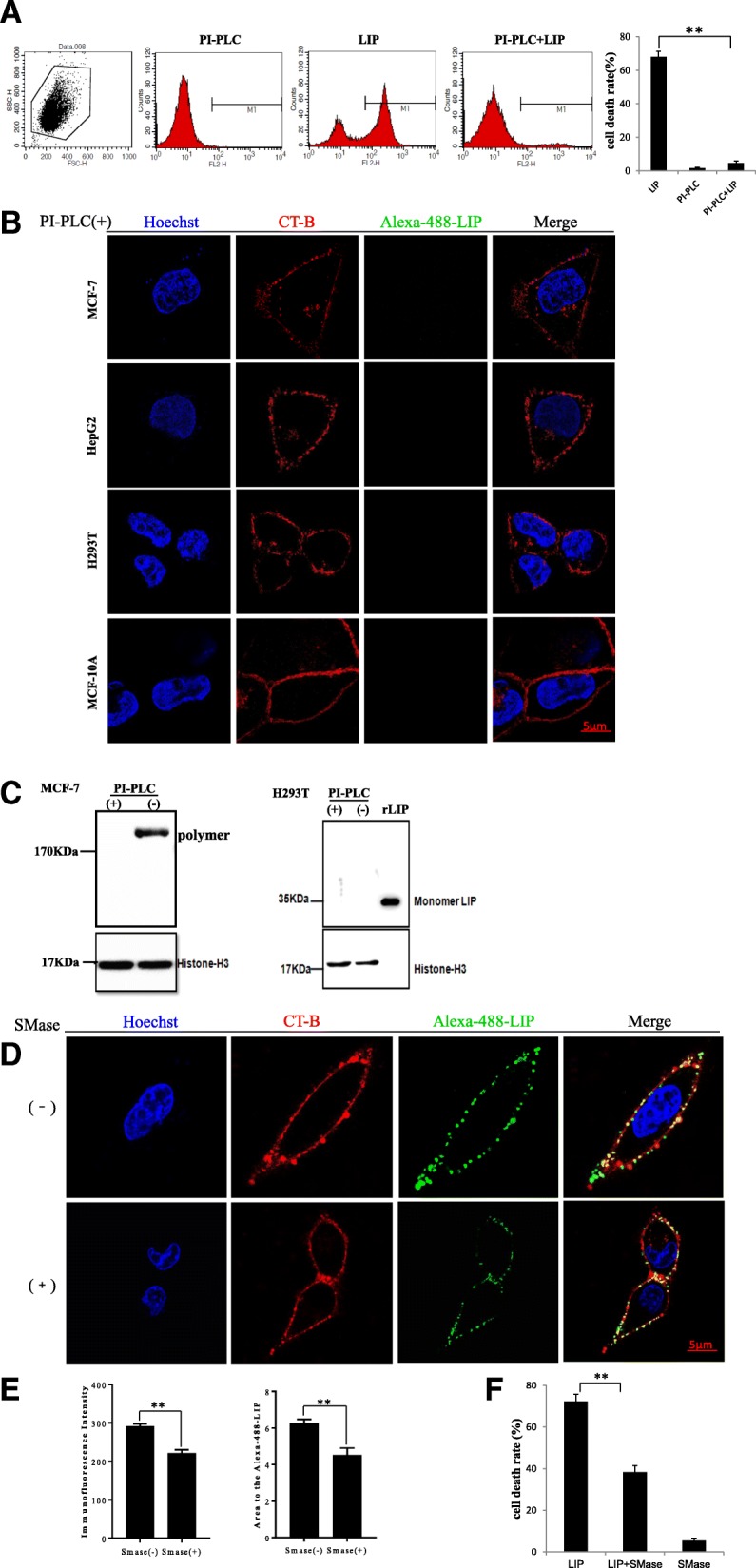
Background: In previous research, we found that lamprey immune protein (LIP) possessed cytocidal activity against tumor cells, but the mechanism of the selective recognition and killing of tumor cells by LIP was not identified.

Methods: Superresolution microscopy, crystallographic structural analysis, glycan chip assay, SPR experiments, FACS assays, computational studies and mass spectrometric analysis firmly establish the mode of action of LIP, which involves dual selective recognition and efficient binding.

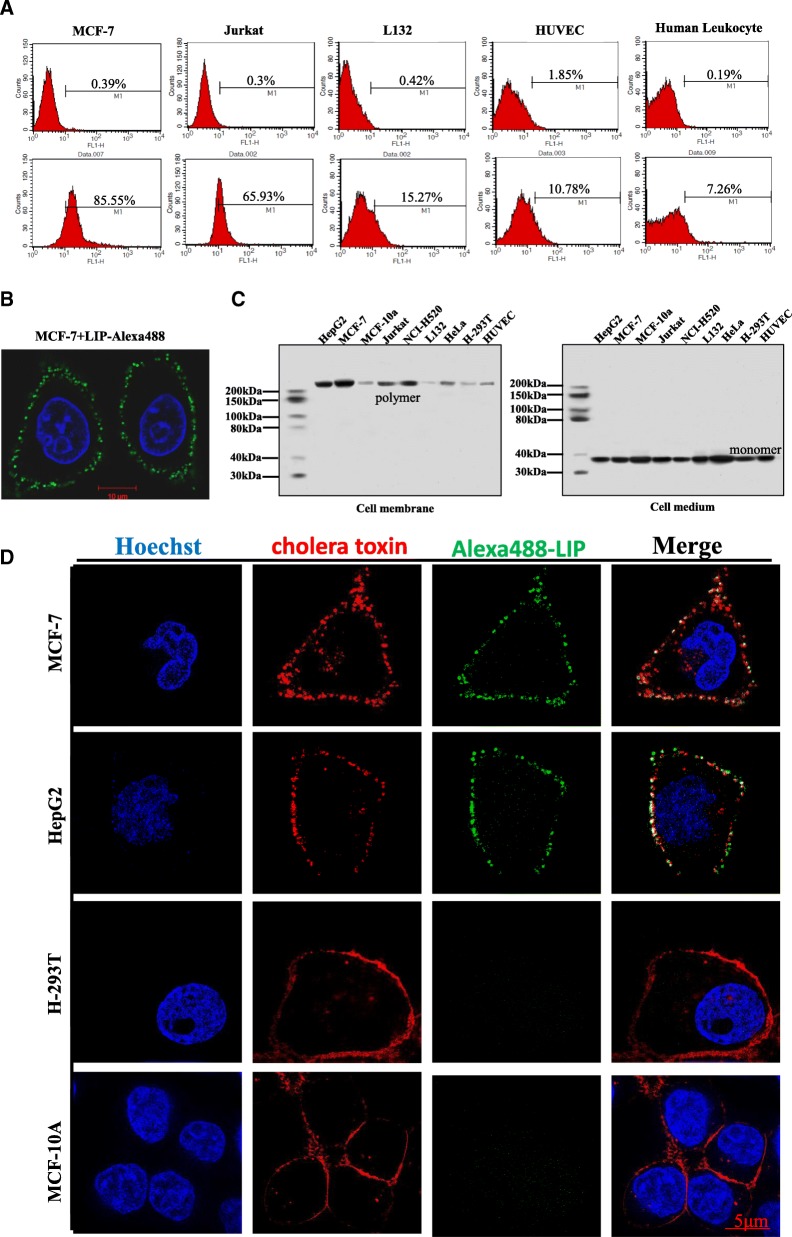
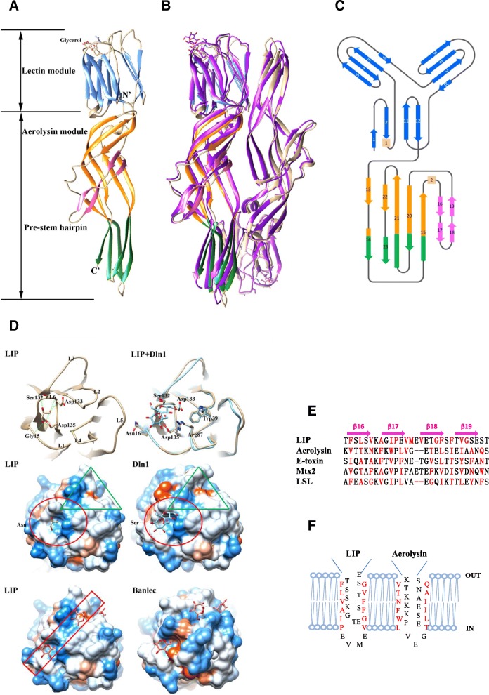
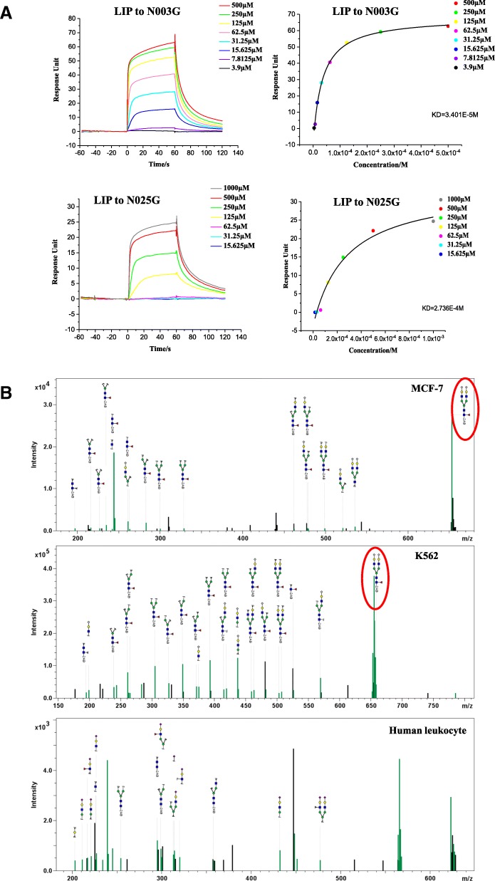
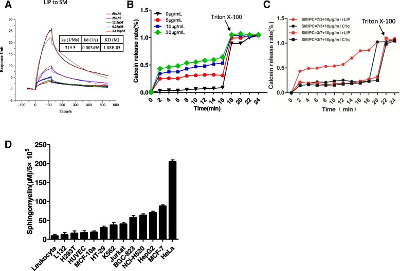
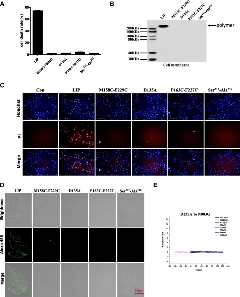
Results: We determined the overall crystallographic structure of LIP at a resolution of 2.25 Å. LIP exhibits an elongated structure with dimensions of 105 Å × 30 Å × 30 Å containing an N-terminal lectin module and a C-terminal aerolysin module. Moreover, the Phe209-Gly232 region is predicted to insert into the lipid bilayer to form a transmembrane β-barrel, in which the hydrophobic residues face the lipid bilayer, and the polar residues constitute the hydrophilic lumen of the pore. We found that LIP is able to kill various human cancer cells with minimal effects on normal cells. Notably, by coupling biochemical and computational studies, we propose a hypothetical mechanism that involves dual selective recognition and efficient binding dependent on both N-linked glycans on GPI-anchored proteins (GPI-APs) and sphingomyelin (SM) in lipid rafts. Furthermore, specific binding of the lectin module with biantennary bisialylated nonfucosylated N-glycan or sialyl Lewis X-containing glycan structures on GPI-APs triggers substantial conformational changes in the aerolysin module, which interacts with SM, ultimately resulting in the formation of a membrane-bound oligomer in lipid rafts.

Conclusions: LIP holds great potential for the application of a marine protein towards targeted cancer therapy and early diagnosis in humans.

Keywords: Cytotoxic activity; GPI-APs; LIP; Lamprey; Selective recognition.


Conflict of interest statement

The authors declare that they have no competing interests.

Figures

Fig. 1
Fig. 1
Selective cytocidal activity of LIP in vitro. (a) Morphological alterations of cells induced by LIP. The cells were incubated with 1 μg/mL LIP at 37 °C for 24 h, observed with a phase-contrast microscope and photographed. (b) Cytocidal activity of LIP against cultured cancer cell lines and normal cells. A total of 5 × 104 cells were preincubated at 37 °C for 20 h and then treated with LIP (final concentration, 1 μg/mL) for 24 h at 37 °C. The cytotoxic activity of LIP was determined using the LDH Cytotoxicity Detection Kit. Each histogram represents the average value of triplicate experiments. Means ± SDs are shown
Fig. 2
Fig. 2
Localization of LIP in the lipid raft microdomains of cancer cell membranes. (a) A total of 5 × 104 cancer cells or normal cells were incubated with Alexa488-tagged LIP (1 μg/mL) at 37 °C for 30 min and then subjected to flow cytometric analysis. The upper panels and lower panels show the results before and after LIP treatment, respectively. (b) The cells were observed and photographed using a Zeiss LSM 780 inverted microscope (magnification: 63×). (c) The cells were incubated with LIP (1 μg/mL) at 37 °C for 30 min. The cell membranes and culture medium were independently collected, resolved by SDS-PAGE and probed by western blotting using anti-LIP antibodies. (d) MCF-7, HepG2, H293T and MCF-10A cells were stained with Alexa555-cholera toxin subunit B (CT-B) prior to staining with Alexa488-tagged LIP. The CT-B was used following the instructions from Thermo Fisher Scientific. The cells were observed and photographed by 3D-SIM superresolution microscopy
Fig. 3
Fig. 3
Overall structure of the LIP dimer. (a) Overall structure of LIP. The lectin module and the middle and C-terminal moieties of the aerolysin module of the LIP subunit are shown in blue, orange, and green, respectively. The prestem hairpin (the putative transmembrane region) is shown in pink. The N and C termini as well as the bound glycerol are labeled. (b) Superimposition of LIP with Dln1. The second subunit of LIP in the asymmetric unit is shown in sandy brown, and Dln1 is shown in purple. (c) Topology diagram of the LIP monomer. (d) Comparison of the ligand-binding sites of LIP (sandy brown) and Dln1 (blue). The interactions of the bound glycerol molecule and residues Gly15, Ser132, Asp133 and Asp135 of the lectin module in LIP are presented (upper panel). The lectin modules of LIP and Dln1 are superimposed. The interactions of the bound sucrose and the residues in the pocket in Dln1 are labeled, as are the corresponding residues in LIP (upper panel). The hydrophobicity of the surfaces of LIP and Dln1 is depicted according to the Kyte-Doolittle scale with colors ranging from Dodger blue for the most hydrophilic to white at 0.0 and orange-red for the most hydrophobic [33] (middle panel). The red circles are the ligand-binding sites. These sites are almost identical, except for the part on the left, which is an asparagine (Asn) in LIP but a serine (Ser) in Dln1. The green triangle is the channel extending from the binding site. Surface representations of LIP and Banlec (lower panel). Banlec and LIP are superimposed, and the ligands from Banlec are presented with the surface of LIP. The belt-shaped channel is marked with a red rectangle. (e) Multiple-sequence alignment of the putative transmembrane region from different aerolysin members. The members include LIP (Lampetra japonica), aerolysin (Aeromonas sobria), E-toxin (Clostridium perfringens), Mtx2 (Lysinibacillus sphaericus) and LSL (Laetiporus sulphureus). Alignments were generated based on the alternating patterns of polar and hydrophobic residues. The hydrophilic residues (facing the pore lumen) and hydrophobic residues (facing the lipid bilayer) are marked in black and red, respectively. (f) Schematic representation of the antiparallel strands forming the β-barrel of LIP and the corresponding residues of aerolysin. The alignment is based on previous reports [21] and sequence similarity. The residues are depicted either facing the lipid bilayer or lining the lumen of the pore
Fig. 4
Fig. 4
Sialylated antennary N-glycan specificity of LIP. (a) BIAcore diagram and saturation curve of LIP bound to N003G and N025G. LIP binds to N003G and N025G with similar low affinities and rapid kinetics. Response units were plotted against protein concentrations. The KD values were calculated by BIAcore T200 analysis software (BIAevaluation version 3.0). (b) The MS/MS spectrum of the glycan from the PI-PLC-treated aqueous fraction after Triton X-114 phase separation from MCF-7 and K562 cells and human leukocytes
Fig. 5
Fig. 5
The cytocidal activity of LIP against tumor cells disappeared upon PI-PLC or SMase treatment. (a) MCF-7 cells were incubated with (+) or without (−) PI-PLC (5 U/mL) for 2 h and then incubated with LIP and stained with PI for flow cytometric analysis. Histogram showing statistics of the above results (right pane). Means ± SDs are shown (n = 3 per group). (b) MCF-7, HepG2, H293T and MCF-10A cells were pretreated with PI-PLC and then stained with Alexa555-cholera toxin subunit B (CT-B) prior to staining with Alexa488-tagged LIP. The cells were observed and photographed by 3D-SIM superresolution microscopy. (c) MCF-7 cells were incubated with (+) or without (−) PI-PLC prior to incubation with LIP. After the cells were washed to remove free LIP, the proteins were separated by SDS-PAGE and detected by immunoblotting with anti-LIP antibodies (left panel). Immunoblotting of proteins in H293T cells incubated with LIP (right panel). (d) MCF-7 cells were pretreated with SMase and then stained with Alexa555-cholera toxin subunit B (CT-B) prior to staining with Alexa488-tagged LIP. (e) The mean immunofluorescence intensity, which was measured as the average gray level, and the area ratio of the Alexa488-tagged  LIP area were examined using Image Pro Plus 6.0. (f) After the preincubation of MCF-7 cells in the presence (+) or absence (−) of SMase, the cells were treated with LIP. Cell death rates were analyzed by the LDH method. Each histogram represents the average value of triplicate experiments (**P < 0.01). Means ± SDs are shown
Fig. 6
Fig. 6
Correlation between the SM-binding ability and cytotoxic activity of LIP. (a) BIAcore diagram and saturation curve of LIP bound to SM. LIP binds to SM with similar low affinity and slow kinetics. The KD values were calculated by BIAcore T200 analysis software (BIAevaluation version 3.0). (b) Dose-dependent effect of LIP on a liposome membrane composed of a mixture of PC and SM (PC:SM = 1:1). (c) The effect of LIP on a liposome membrane composed of a mixture of PC and SM depends on the SM content (PC:SM = 3:7, PC:SM = 7:3). Recombinant L-C1q proteins were used as a negative control in these experiments. (d) The content of sphingomyelin in different types of cells
Fig. 7
Fig. 7
Dual recognition mode of LIP for cancer cells. (a) Binding mode of LIP with the disaccharides of Neu5Gc coupled with 2,6-galactose and 2,3-galactose at the N-terminal domain. The disaccharide and the key residues that interacted with it are shown in sticks and colored yellow and green, respectively. The LIP protein is shown in a cartoon representation in green. (b) Binding mode of LIP with SM at the C-terminal module. SM is shown as rainbow spheres. LIP protein is shown in cartoon and surface representations and colored cyan. The key residues that interacted with SM are labeled, shown in stick representation and colored green. (c) Fluorescence spectra of LIP under different conditions. The LIP stock solutions and N003G or SM stock solutions, respectively, were mixed in phosphate buffer. The resultant mixture was equilibrated for 2 min before recording the steady-state fluorescence spectrum, and the emission spectra were obtained at wavelengths ranging from 290 to 495 nm. Values are the means of five independent experiments. (d) Rapid kinetics of the binding of N003G to LIP. Stopped-flow fluorescence measurements of the binding of N003G to LIP. The experiment was performed in PBS at 25 °C. All data sets were analyzed simultaneously with proper weighting to yield best-fit parameters. K1 = 24.925540 s− 1, K2 = 2.128309 ± 0.055980 s− 1, K3 = -0.0063 s− 1
Fig. 8
Fig. 8
Mutagenesis confirms the key residues. (a) MCF-7 cells were treated with LIP or other mutants. MCF-7 cells were plated in 96-well plates at a density of 5 × 104 cells/well and treated with 1 μg/mL LIP and mutants at 37 °C for 24 h. Cell death rates were analyzed by the LDH method. Each histogram represents the average value of triplicate experiments (**P < 0.01). Means ± SDs are shown. (b) Immunoblot analysis of LIP and mutants after incubation with MCF-7 cells. The observed bands represent the cell-bound protein from cell membranes. The band corresponding to polymer is indicated with black arrows. (c) Staining dead cells with propidium iodide (PI) for high content screening (magnification: 40×). MCF-7 cells were plated in 96-well plates at a density of 5 × 104 cells/well and treated with 1 μg/mL LIP and mutants for 24 h. Cells were washed twice with phosphate-buffered saline (PBS) and stained with PI and Hoechst (Sigma) for 20 min to visualize the cell nuclei. The samples were analyzed on a High Content Screen (PerkinElmer, USA). (d) Binding of Alexa488-labeled LIP and mutants to MCF-7 cells. (e) BIAcore diagrams of the binding of the D135A mutant of LIP to N003G. The D135A mutant abolished the binding to N003G

Similar articles

Cited by

References

    1. Bischofberger M, Gonzalez MR, van der Goot FG. Membrane injury by pore-forming proteins. Curr Opin Cell Biol. 2009;21:589–595. doi: 10.1016/j.ceb.2009.04.003. - DOI - PubMed
    1. Sousa MV, Richardson M, Fontes W, Morhy L. Homology between the seed cytolysin enterolobin and bacterial aerolysins. J Protein Chem. 1994;13:659–667. doi: 10.1007/BF01886950. - DOI - PubMed
    1. Mancheño JM, Tateno H, Goldstein IJ, Martínez-Ripoll M, Hermoso JA. Structural analysis of the Laetiporus sulphureus hemolytic pore-forming lectin in complex with sugars. J Biol Chem. 2005;280:17251–17259. doi: 10.1074/jbc.M413933200. - DOI - PubMed
    1. Lashuel HA, Lansbury PT., Jr Are amyloid diseases caused by protein aggregates that mimic bacterial pore-forming toxins? Q Rev Biophys. 2006;39:167–201. doi: 10.1017/S0033583506004422. - DOI - PubMed
    1. Saur M, Moeller V, Kapetanopoulos K, Braukmann S, Gebauer W, Tenzer S, Markl J. Acetylcholine-binding protein in the hemolymph of the planorbid snail Biomphalaria glabrata is a pentagonal dodecahedron (60 subunits) PLoS One. 2012;7:e43685. doi: 10.1371/journal.pone.0043685. - DOI - PMC - PubMed

Publication types

LinkOut - more resources