. 2019 May 27;38(1):221.
doi: 10.1186/s13046-019-1226-8.

Inhibition of ERK1/2 in cancer-associated pancreatic stellate cells suppresses cancer-stromal interaction and metastasis

Affiliations

Inhibition of ERK1/2 in cancer-associated pancreatic stellate cells suppresses cancer-stromal interaction and metastasis

Zilong Yan et al. J Exp Clin Cancer Res. .

Abstract

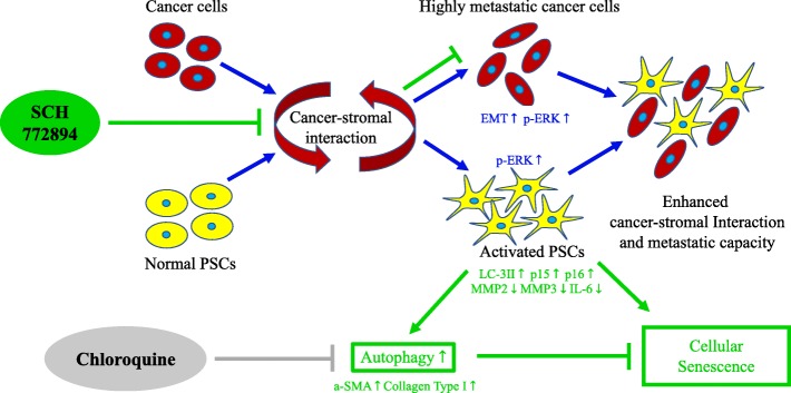
Background: Extracellular signal-regulated kinases (ERKs) have been related to multiple cancers, including breast cancer, hepatocellular cancer, lung cancer and colorectal cancer. ERK1/2 inhibitor can suppress growth of KRAS-mutant pancreatic tumors by targeting cancer cell. However, no studies have shown the expression of ERK1/2 on pancreatic stromal and its effect on pancreatic cancer-stromal interaction.

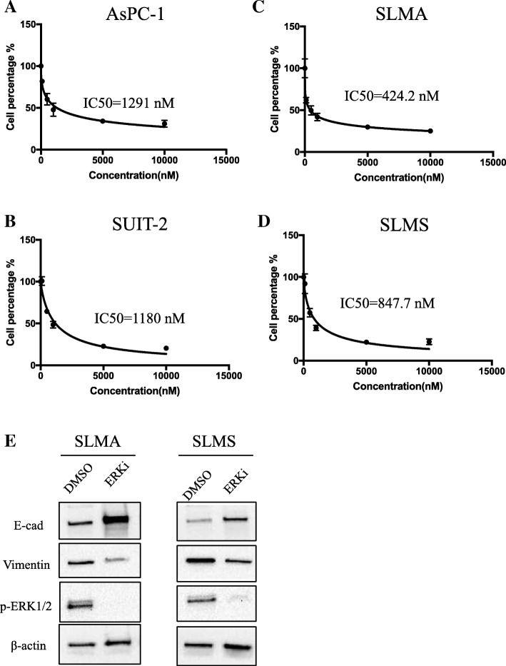
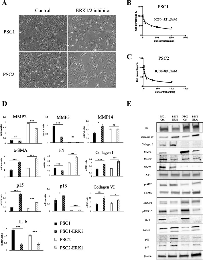
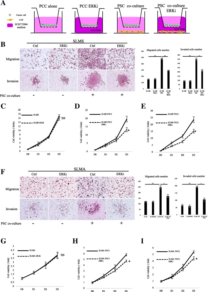
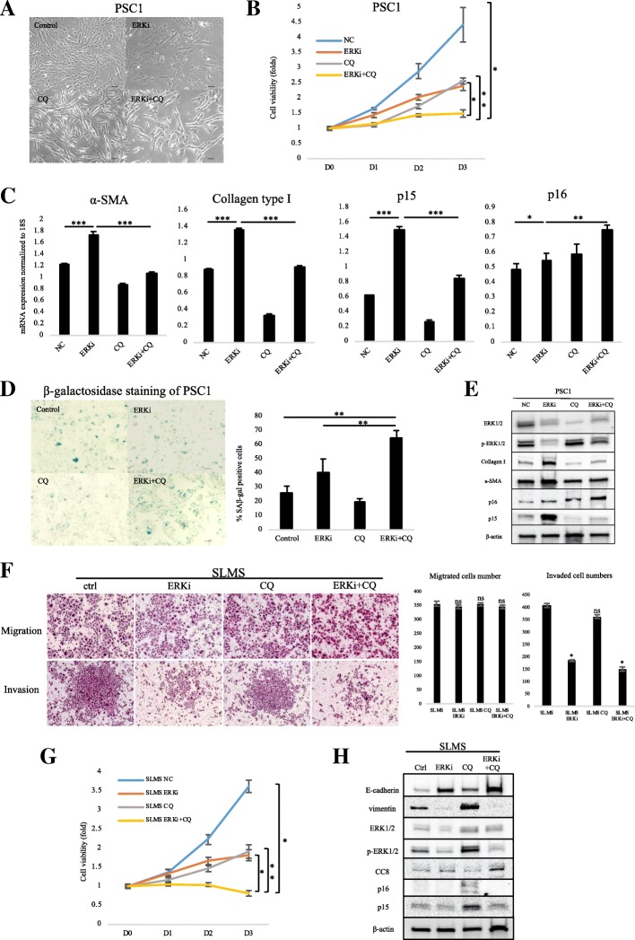
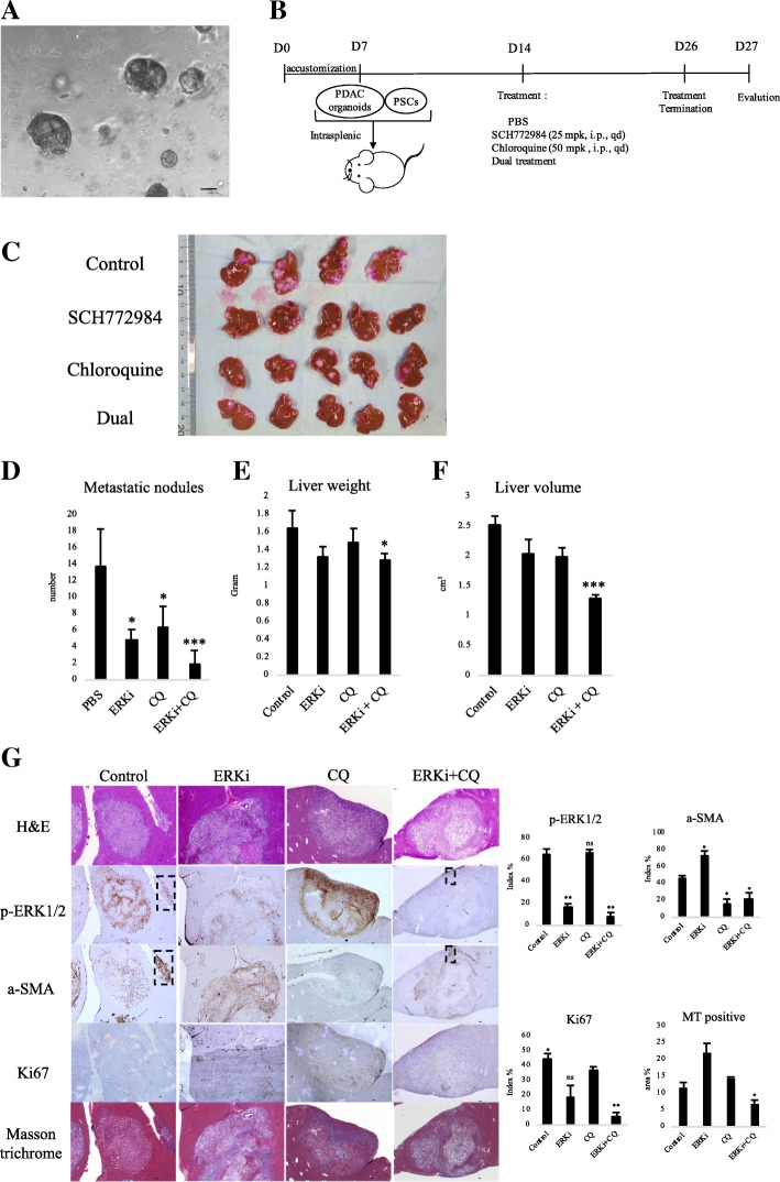
Methods: Immunohistochemistry and western blotting were performed to detect the expression of p-ERK1/2 in pancreatic tissues and cells. Cell viability assay was used to study IC50 of ERK inhibitor on pancreatic cancer cells (PCCs) and primary cancer-associated pancreatic stellate cells (PSCs). Transwell migration, invasion, cell viability assay, senescence β-galactosidase staining were performed to determine the effect of ERK inhibitor on PCCs and PSCs in vitro and in vivo. The expression of key factors involved in autophagy and epithelial-to-mesenchymal transition (EMT) process were evaluated by western blotting. The expression of key factors related to cell invasiveness and malignancy were confirmed by qRT-PCR. Co-transplantation of PCC Organoid and PSC using a splenic xenograft mouse model was used to evaluated combined treatment of ERK inhibitor and autophagy inhibitor.

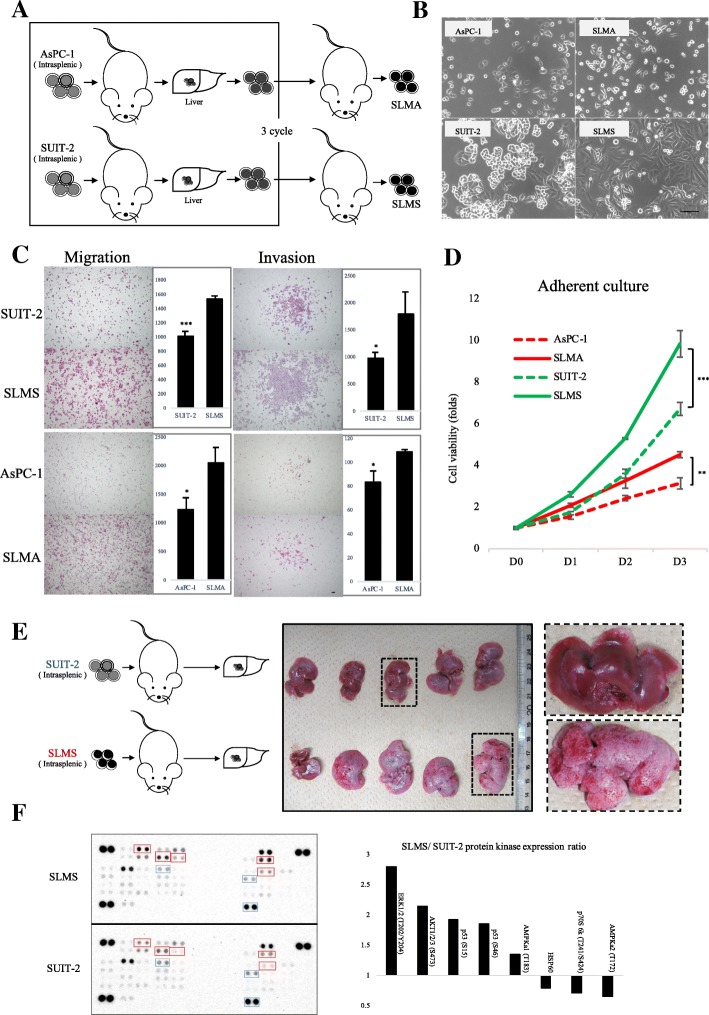
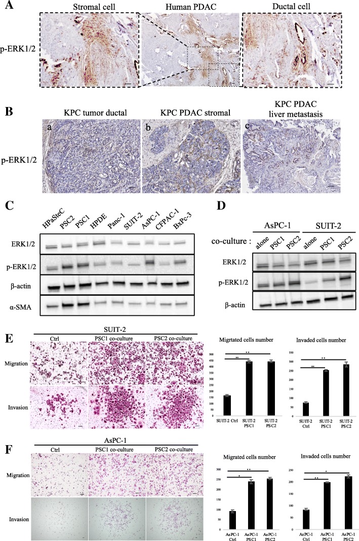
Results: Immunohistochemical staining in pancreatic tumor samples and transgenetic mice detected p-ERK1/2 expression in both cancer cells and stromal cells. In pancreatic tissues, p-ERK1/2 was strongly expressed in cancer-associated PSCs compared with cancer cells and normal PSCs. PSCs were also significantly more sensitive to ERK1/2 inhibitor treatment. Inhibition of ERK1/2 suppressed EMT transition in HMPCCs, upregulated cellular senescence markers, activated autophagy in cancer-associated PSCs; and suppressed cancer-stromal interaction, which enhanced invasiveness and viability of cancer cells. We also found that chloroquine, an autophagy inhibitor, suppressed ERK inhibition-induced autophagy and promoted PSC cellular senescence, leading to significantly decreased cell proliferation. The combination of an ERK inhibitor and autophagy inhibitor suppressed liver metastasis in a splenic pancreatic cancer organoid xenograft mouse model.

Conclusions: These data indicate that inhibition of ERK1/2 in cancer-associated pancreatic stellate cells suppresses cancer-stromal interaction and metastasis.

Keywords: Cancer–stromal interaction; Cellular senescence; ERK1/2; Pancreatic cancer; Pancreatic stellate cell.


Conflict of interest statement

The authors declare that they have no competing interests.

Figures

Fig. 1
Fig. 1
Establishment of highly metastatic PCCs. a Establishing PDAC cells. Parental PDAC cells were splenic transplanted into nude mice; liver metastases were harvested after 2–4 weeks. This process was performed 3 times. b Cellular morphology of parental PDAC cells and highly metastatic PDAC cells. Scale bars = 100 μm. c Migration and invasion assays were performed over 36 and 18 h respectively. Graphs show numbers of cells calculated from five fields. Original magnification: 40×. Scale bars =100 μm. *P < 0.05, ***P < 0.001. d Cell viability of cancer cells as determined by CellTiter-Glo luminescent cell viability assay. *P < 0.05, **P < 0.01. e SUIT-2 and SLMS cells were intrasplenic injected in nude mice and the liver metastases were harvested. Gross pathology indicated metastatic lesions. f Phospho-protein kinase array of SUIT-2 and SLMS cells. Right: most significant gene alterations
Fig. 2
Fig. 2
Expression of p-ERK1/2 in PDAC tissues and pancreatic cell lines. a p-ERK1/2 expression was detected in both pancreatic cancer cells and stromal cells. Scale bars =100 μm. b p-ERK1/2 expression was detected in KPC mice cancer cells (a) and stromal cells (b) of pancreatic primary tumor, and liver metastases (c). Scale bars =100 μm. c Western blot of ERK1/2, p-ERK1/2, and α-SMA levels in pancreatic cells. d Western blot of ERK1/2 and p-ERK1/2 levels in PCCs, alone or after co-culture with PSCs. PCCs were seeded in 24-well plates while PSCs were seeded in the upper transwell chamber with 3-μm pore size. e SUIT-2, (f) AsPC-1 Migration and invasion assays were performed for 18 and 36 h, respectively. PSCs were seeded in 24-well plates while PCCs were seeded in the upper transwell chamber of 8-μm pore size. Graphs show numbers of cells calculated from five fields. Scale bars =100 μm. *P < 0.05, **P < 0.01
Fig. 3
Fig. 3
Inhibition of ERK1/2 decreased PDAC cell viability and EMT transition. a AsPC-1, (b) SUIT-2, (c) SLMA, and (d) SLMS cell viability after 72 h; treatment with various concentrations of ERK inhibitor after. IC50 values are indicated. e Western blot of E-cadherin, vimentin, and p-ERK1/2 levels of highly metastatic cancer cells after treatment with ERK inhibitor SCH772984 at IC50 value. The indicated protein was extracted exclusively from the living adherent cells. Negative control: DMSO
Fig. 4
Fig. 4
Inhibition of ERK1/2 facilitated PSCs atrophy and induces p16, α-SMA. a Microphotograph of PSCs after treatment with DMSO and/or ERK inhibitor. Scale bars = 100 μm. b Viability of PSC1 and (c) PSC2 cells, as determined by CellTiter-Glo luminescent cell viability assay after 72 h’ treatment with indicated concentrations of ERK inhibitor; IC50 values are indicated. d qRT-PCR of PSCs shows mRNA expression changes after ERK inhibitor treatment. *P < 0.05, **P < 0.01, ***P < 0.001. e The indicated protein levels of PSCs were evaluated after treatment with DMSO as control and/or ERK inhibitor SCH772984 at IC50 value
Fig. 5
Fig. 5
Inhibition of ERK1/2 suppressed PCC-PSC interaction by preferentially targeting PSC. a In indirect co-culture experiments, first PSCs were seeded, and 24 h later, medium was replaced and transwell chambers (8-μm pores; Becton Dickinson) were placed in 24-well dishes, and then PCCs were seeded into the transwell chambers. After incubation for the indicated time, migration and invasion were evaluated by counting the cells that had invaded to the lower chamber. SCH772984 dose was used IC50 value of PSC2 cells, 89 nM. b Migration and invasion assays were performed for 18 and 36 h, respectively. Graphs show numbers of cells calculated from five fields. Scale bars =100 μm. *P < 0.05, **P < 0.01. c Viability of SLMS cells co-cultured with (d) PSC1 or (e) PSC2 cells after DMSO or SCH772984 treatment; (g) Viability of SLMA cells co-cultured with (h) PSC1 or (i) PSC2 cells after DMSO or SCH772984 treatment was determined by CellTiter-Glo luminescent cell viability assay. SCH772984 dose was used IC50 value of PSC1, 321 nM; and PSC2, 90 nM. *P < 0.05. Columns, mean fold changes of three experiments done in triplicate
Fig. 6
Fig. 6
Dual treatment of SCH772984 and CQ decreased cell viability and induced senescence of PSCs. a Western blot of fibronectin, α-SMA, LC3-II, Akt and p-Akt levels of PSCs after treatment with ERK inhibitor. b Microphotograph of PSCs after indicated agent treatment. Scale bars = 100 μm. c Cell viability of PSCs after indicated agent treatment. d qRT-PCR shows mRNA expression of α-SMA, Collagen Type I, p15 and p16. *P < 0.05, **P < 0.01, ***P < 0.001. e β-galactosidase staining of PSCs after indicated agent treatment. Bottom: graphs show the quantification of β-gal-positive cells calculated from five fields. Scale bars =100 μm. *P < 0.05. e Migration and invasion assays were performed for 18 and 36 h, respectively. Graphs show numbers of cells calculated from five fields. Scale bars =100 μm. *P < 0.05. f Cell viability of PCCs after indicated agent treatment. Columns, mean fold changes in three experiments done in triplicate. g Western blot of indicated protein levels in PCCs after indicated treatment
Fig. 7
Fig. 7
Dual treatment of SCH772984 and CQ decreased liver metastasis in xenograft organoid model with PSC co-transplantation. a Microphotograph of KPC mouse-derived cancer organoid. Scale bars =100 μm. b Scheme of xenograft experiment. Female nude mice were intrasplenic transplanted with cancer organoids with PSCs and randomized divided into four groups (n = 5/group). One week after implantation, mice were dosed once daily with vehicle, SCH772984 (25 mg/kg), Chloroquine (50 mg/kg), or dual treatment of each group for 13 days. Dosing occurred from day 14 to day 26. At day 27, mice were sacrificed and liver metastases were harvested. c Gross pathology showed significantly reduced liver metastasis formation after dual treatment of SCH772984 with chloroquine. d Liver metastasis nodules were significantly reduced in samples treated with SCH772984 or CQ, or both. *P < 0.05, ***P < 0.001. e Tumor weight and (f) volume were significantly decreased only in the dual-treatment group. *P < 0.05, ***P < 0.001. g Immunohistochemical staining shows decreased p-ERK1/2 expression in SCH772984 group, and decreased α-SMA expression in CQ group; The dual-treated group showed significant reductions of p-ERK1/2, α-SMA, Ki67 and collagen fibers. Corresponding rectangles indicated a-SMA-positive PSCs. Right: quantification of gene expression from five fields. Scale bars =100 μm. *P < 0.05, **P < 0.01
Fig. 8
Fig. 8
Mechanism of p-ERK1/2 inhibition on cancer–stromal interaction. In PDAC progression, interaction between cancer cells and stromal cells is a key regulator of ERK1/2 activation, during which cancer cells transform to an ERK1/2 activation phenotype and exhibit EMT transition tendency, and PSCs turn activated from their quiescent status. Thus enhancement of cancer–stromal interactions result in greater metastatic capacity; Whereas SCH772984 suppressed EMT transition of cancer cells, and upregulated senescence markers p15 and p16, malignancy-related genes MMP2, MMP3 and IL-6, and fibrosis markers α-SMA and Collagen Type I in PSCs. Also, its combination with an autophagy inhibitor, chloroquine, suppresses SCH772984-induced autophagy. Therefore, the combination therapy possibly leads to strong induction of cellular senescence in PSCs. In summary, dual treatment with ERK inhibitor and CQ inhibit cancer–stromal interaction and metastatic capacity

Similar articles

Cited by

References

    1. Stewart BW, Wild CP. World cancer report 2014. World Heal Organ [Internet] 2014;1–2. Available from: https://www.who.int/cancer/publications/WRC_2014/en/.
    1. Hess KR, Varadhachary GR, Taylor SH, Wei W, Raber MN, Lenzi R, et al. Metastatic patterns in adenocarcinoma. Cancer. 2006;106:1624–1633. doi: 10.1002/cncr.21778. - DOI - PubMed
    1. Howlader N, Noone AM, Krapcho M et al. SEER Cancer Statistics Review, 1975–2013. Natl Cancer Institute Bethesda, MD [Internet]. 2016. Available from: http://seer.cancer.gov/csr/1975_2013/.
    1. Gleisner AL, Assumpcao L, Cameron JL, Wolfgang CL, Choti MA, Herman JM, et al. Is resection of periampullary or pancreatic adenocarcinoma with synchronous hepatic metastasis justified? Cancer. 2007;110:2484–2492. doi: 10.1002/cncr.23074. - DOI - PubMed
    1. Neesse A, Michl P, Frese KK, Feig C, Cook N, Jacobetz MA, et al. Stromal biology and therapy in pancreatic cancer. Gut. 2011. - PubMed

MeSH terms