. 2019 May 27;9(1):16.
doi: 10.1186/s13395-019-0197-y.

A GDF11/myostatin inhibitor, GDF11 propeptide-Fc, increases skeletal muscle mass and improves muscle strength in dystrophic mdx mice

Affiliations

A GDF11/myostatin inhibitor, GDF11 propeptide-Fc, increases skeletal muscle mass and improves muscle strength in dystrophic mdx mice

Quan Jin et al. Skelet Muscle. .

Abstract

Background: Growth differentiation factor 11 (GDF11) is a member of the transforming growth factor β superfamily. The GDF11 propeptide, which is derived from the GDF11 precursor protein, blocks the activity of GDF11 and its homolog, myostatin, which are both potent inhibitors of muscle growth. Thus, treatment with GDF11 propeptide may be a potential therapeutic strategy for diseases associated with muscle atrophy like sarcopenia and the muscular dystrophies. Here, we evaluate the impact of GDF11 propeptide-Fc (GDF11PRO-Fc) gene delivery on skeletal muscle in normal and dystrophic adult mice.

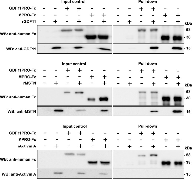
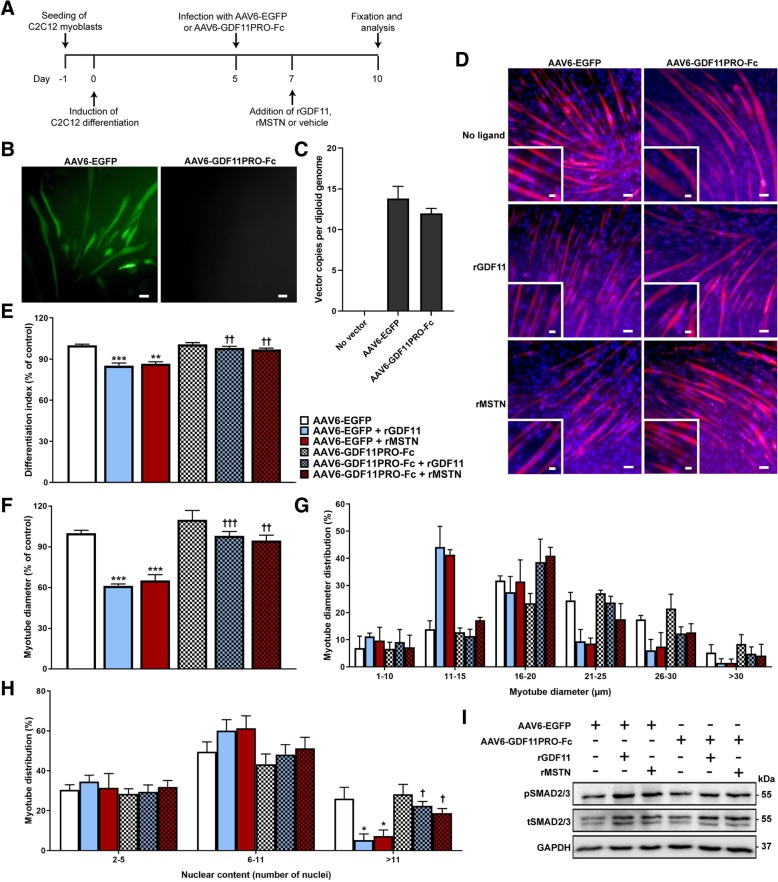
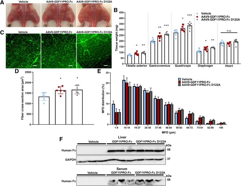
Methods: A pull-down assay was used to obtain physical confirmation of a protein-protein interaction between GDF11PRO-Fc and GDF11 or myostatin. Next, differentiated C2C12 myotubes were treated with AAV6-GDF11PRO-Fc and challenged with GDF11 or myostatin to determine if GDF11PRO-Fc could block GDF11/myostatin-induced myotube atrophy. Localized expression of GDF11PRO-Fc was evaluated via a unilateral intramuscular injection of AAV9-GDF11PRO-Fc into the hindlimb of C57BL/6J mice. In mdx mice, intravenous injection of AAV9-GDF11PRO-Fc was used to achieve systemic expression. The impact of GDF11PRO-Fc on muscle mass, function, and pathological features were assessed.

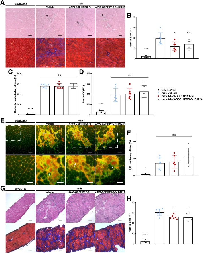
Results: GDF11PRO-Fc was observed to bind both GDF11 and myostatin. In C2C12 myotubes, expression of GDF11PRO-Fc was able to mitigate GDF11/myostatin-induced atrophy. Following intramuscular injection in C57BL/6J mice, increased grip strength and localized muscle hypertrophy were observed in the injected hindlimb after 10 weeks. In mdx mice, systemic expression of GDF11PRO-Fc resulted in skeletal muscle hypertrophy without a significant change in cardiac mass after 12 weeks. In addition, grip strength and rotarod latency time were improved. Intramuscular fibrosis was also reduced in treated mdx mice; however, there was no change seen in central nucleation, membrane permeability to serum IgG or serum creatine kinase levels.

Conclusions: GDF11PRO-Fc induces skeletal muscle hypertrophy and improvements in muscle strength via inhibition of GDF11/myostatin signaling. However, GDF11PRO-Fc does not significantly improve the dystrophic pathology in mdx mice.

Keywords: AAV; Duchenne muscular dystrophy; GDF11; Gene delivery; Gene therapy; Hypertrophy; Myostatin; mdx.


Conflict of interest statement

The authors declare that they have no competing interests.

Figures

Fig. 1
Fig. 1
GDF11PRO-Fc associates with GDF11 and MSTN. Protein-protein interactions between GDF11PRO-Fc or MPRO-Fc and rGDF11, rMSTN, or rActivin A were determined by a pull-down assay. GDF11PRO-Fc or MPRO-Fc was incubated with rGDF11, rMSTN, or rActivin A for 1 h at 4 °C. Fc-fused protein complexes were separated on a protein A/G-coated agarose resin and eluates were run on a 12% SDS-PAGE gel under reducing conditions and probed by western blot. Input control was 5% of the input material. WB western blot
Fig. 2
Fig. 2
GDF11PRO-Fc blocks GDF11/MSTN-induced myotube atrophy in C2C12 cells. a Schematic detailing experimental timeline in C2C12 myotubes. AAV6-EGFP or AAV6-GDF11PRO-Fc was added to C2C12 myotubes at a MOI of 105 on day 5 post-differentiation and 100 ng/ml rGDF11 or rMSTN was added on day 7. Myotubes were stained and analyzed on day 10. b EGFP expression was evident at 48–72 h in C2C12 myotubes treated with AAV6-EGFP (MOI 10 [5]). Scale bars represents 50 μm. c Vector genome copy number per diploid genome in C2C12 myotubes 72 h after addition of AAV6-EGFP or AAV6-GDF11PRO-Fc (MOI 10 [5]). d Representative immunofluorescence images of C2C12 myotubes. C2C12 myotube membranes were visualized by staining with an anti-dystrophin antibody (red). Nuclei were stained with DAPI (blue). Inset shows a zoomed-in region. Scale bars represent 50 μm (main panel) and 25 μm (panel inset). e The fraction of nuclei incorporated into myotubes (differentiation index) was calculated and presented as a percentage of control. f Average myotube diameter relative to control and (g) distribution of diameter measurements. For myotube diameter measurements, each myotube was measured at three points along the length of the myotube and averaged. h Number of nuclei incorporated per myotube. A minimum of 50 myotubes were analyzed per experimental condition. i pSMAD2/3 relative to tSMAD2/3 was assessed by western blot. Equal protein loading was verified by Ponceau S staining and GAPDH was used as a loading control. Data represents results from three separate experiments. All error bars represent mean ± SEM. *p < 0.05; **p < 0.01; ***p < 0.001; n.s. not significant; compared to AAV6-EGFP-treated control. †p < 0.05; ††p < 0.01; †††p < 0.001; compared to AAV6-EGFP + ligand-treated. pSMAD2/3: phosphorylated SMAD2/3; tSMAD2/3: total SMAD2/3
Fig. 3
Fig. 3
GDF11PRO-Fc induces localized skeletal muscle hypertrophy after intramuscular gene delivery. 8-week-old C57BL/6J mice were treated with AAV9-GDF11PRO-Fc (n = 5) or vehicle (n = 5) via unilateral intramuscular injection into the right-side hindlimb. The contralateral left-side hindlimb was not treated. Mice were euthanized 10 weeks post-treatment. a Average bodyweight over time. b Hindlimb grip strength normalized to bodyweight at 10 weeks post-treatment. Shown are measurements from a single hindlimb and both hindlimbs. c Representative gross hindlimb musculature. The injected hindlimb is designated with a black arrow. d Wet tissue weight of tibialis anterior and gastrocnemius. e Representative immunofluorescence images of injected right-side gastrocnemius cross-sections stained with AlexaFluor-488-conjugated WGA to visualize myofibers. Scale bars represent 100 μm. f Average myofiber cross-section area in the injected right-side gastrocnemius. g Myofiber MFD distribution in the injected right-side gastrocnemius. A minimum of 500 myofibers were measured per mouse. h Western blot identification of GDF11PRO-Fc in tissue lysates from injected right-side gastrocnemius and liver samples of treated mice. GDF11PRO-Fc was not detectable in vehicle-treated mice. Equal protein loading was verified by Ponceau S staining and GAPDH was used as a loading control. All error bars represent mean ± SEM. *p < 0.05; **p < 0.01; n.s. not significant; compared to vehicle-treated control. TA tibialis anterior, Gas gastrocnemius
Fig. 4
Fig. 4
GDF11PRO-Fc improves grip strength and rotarod performance in dystrophic mdx mice. 6-week-old mdx mice were treated with AAV9-GDF11PRO-Fc (n = 7), AAV9-GDF11PRO-Fc D122A (n = 7), or vehicle (n = 7) via tail vein injection. a Average bodyweight over time. b Forelimb grip strength normalized to bodyweight over time. c Best rotarod time-to-fall recorded from pre-treatment (baseline) and 12 weeks post-treatment. The best time from three attempts was used for analysis. d Total distance run on treadmill endurance test from pre-treatment (baseline) and 12 weeks post-treatment. All error bars represent mean ± SEM. *p < 0.05; **p < 0.01; n.s. not significant; compared to vehicle-treated control
Fig. 5
Fig. 5
GDF11PRO-Fc induces skeletal muscle hypertrophy in dystrophic mdx mice. 6-week-old mdx mice were treated with AAV9-GDF11PRO-Fc (n = 7), AAV9-GDF11PRO-Fc D122A (n = 7), or vehicle (n = 7) via tail vein injection. Mice were euthanized 12 weeks post-treatment. a Representative gross hindlimb musculature. b Wet tissue weight of limb muscles, diaphragm, and heart. c Representative immunofluorescence images of gastrocnemius cross-sections stained with AlexaFluor-488-conjugated WGA to visualize myofibers. Scale bars represent 100 μm. d Average myofiber cross-section area in the gastrocnemius. e Myofiber MFD distribution in the gastrocnemius. A minimum of 500 myofibers were measured per mouse. f Identification of GDF11PRO-Fc by western blot in liver tissue lysate and serum of mice treated with AAV9-GDF11PRO-Fc. Equal protein loading was verified by Ponceau S staining and GAPDH was used as a loading control in liver tissue lysates. *p < 0.05; **p < 0.01; ***p < 0.001; n.s. not significant; compared to vehicle-treated control
Fig. 6
Fig. 6
GDF11PRO-Fc does not mitigate the dystrophic pathology in mdx mice. 6-week-old mdx mice were treated with AAV9-GDF11PRO-Fc (n = 7), AAV9-GDF11PRO-Fc D122A (n = 7) or vehicle (n = 7) via tail vein injection. Age-matched C57BL/10J (n = 5) mice were also included as a wild type control. Mice were euthanized 12 weeks post-treatment. a Representative HE and MTC staining from gastrocnemius cross-sections of C57BL/10J and mdx mice. Central nucleation is evident in all the mdx groups. Localized areas of myofiber necrosis and regeneration are marked with a black arrow. Fibrotic area is represented by the blue region on MTC staining. Scale bars represent 50 μm in HE sections and 100 μm in MTC sections. b Fibrotic area in the gastrocnemius expressed as a percentage of the total muscle cross-section area. c Percentage of myofibers exhibiting central nucleation. d Measurement of circulating levels of serum CK, a marker of muscle damage. e Representative immunofluorescence images of mouse IgG staining (red) in gastrocnemius tissue cross-sections. Positive staining for IgG indicates myofiber permeability to serum proteins and loss of membrane integrity. Sections were co-stained with WGA to visualize individual myofibers. Zoomed-in area is marked with a white box. Characteristic IgG-positive myofibers are depicted with a white arrow. Scale bars represent 100 μm. f The area of IgG-positive myofibers expressed as a percentage of the total muscle cross-section area. g Representative HE and MTC staining from diaphragm cross-sections of C57BL/10J and mdx mice. Extensive pathology is apparent in mdx but not C57BL/10J mice. Scale bars represent 100 μm. h Fibrotic area in the diaphragm expressed as a percentage of the total cross-section area. *p < 0.05; ***p < 0.001; ****p < 0.0001; n.s. not significant; compared to vehicle-treated mdx controls

Similar articles

Cited by

References

    1. Nakashima M, Toyono T, Akamine A, Joyner A. Expression of growth/differentiation factor 11, a new member of the BMP/TGFβ superfamily during mouse embryogenesis. Mech Dev. 1999;80:185–189. doi: 10.1016/S0925-4773(98)00205-6. - DOI - PubMed
    1. McPherron AC, Lawler AM, Lee S-J. Regulation of anterior/posterior patterning of the axial skeleton by growth/differentiation factor 11. Nat Genet. 1999;22:260–264. doi: 10.1038/10320. - DOI - PubMed
    1. Kim J, et al. GDF11 controls the timing of progenitor cell competence in developing retina. Science. 2005;308:1927–1930. doi: 10.1126/science.1110175. - DOI - PubMed
    1. Harmon EB, et al. GDF11 modulates NGN3+ islet progenitor cell number and promotes beta-cell differentiation in pancreas development. Dev Camb Engl. 2004;131:6163–6174. - PubMed
    1. Oh SP, et al. Activin type IIA and IIB receptors mediate Gdf11 signaling in axial vertebral patterning. Genes Dev. 2002;16:2749–2754. doi: 10.1101/gad.1021802. - DOI - PMC - PubMed

Publication types

MeSH terms

LinkOut - more resources