. 2019 May 27;9(1):7856.
doi: 10.1038/s41598-019-44289-x.

The transcriptome of Cryptosporidium oocysts and intracellular stages

Affiliations

The transcriptome of Cryptosporidium oocysts and intracellular stages

Lucas V S Matos et al. Sci Rep. .

Abstract

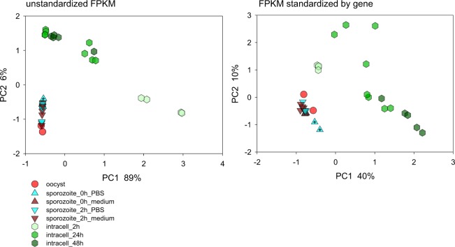
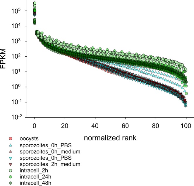
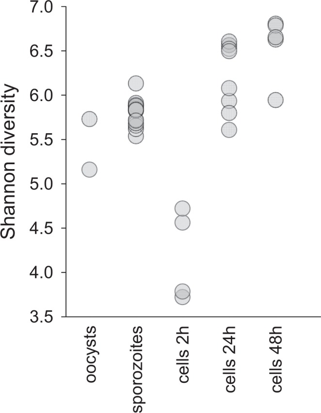
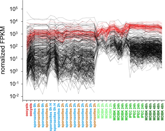
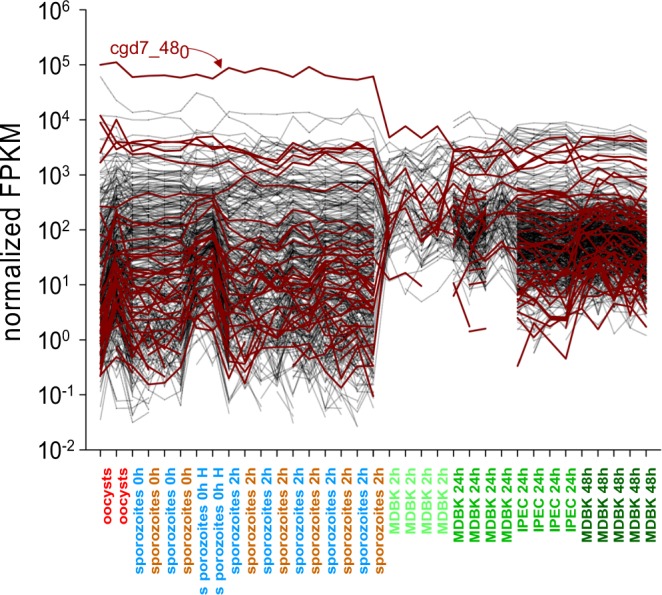
Human cryptosporidiosis is caused primarily by two species of apicomplexan parasites, Cryptosporidium parvum and C. hominis. Although infection of cell monolayers with sporozoites does not support the complete parasite life cycle, the in vitro system is used to study the asexual phase of multiplication, which consists of two generations of merogony. To better understand host-parasite interaction and to gain insight into gene regulatory processes driving the complex life cycle of Cryptosporidium parasites, we analyzed the transcriptome of C. parvum in oocysts, sporozoites and infected cell monolayers 2-48 h post-infection. Analysis of RNA-Seq data from replicate oocyst, sporozoite and intracellular samples revealed significant differences between transcriptomes expressed outside and inside the host cell. Compared to the transcriptome found in the host cell, the oocyst transcriptome is less diverse. Biological processes significantly over-represented intracellularly relate to biosynthetic processes. Genes significantly overexpressed in oocysts show evidence of specialized functions not found in other Apicomplexa. A more comprehensive view of gene regulation during the Cryptosporidium life cycle will require the analysis of later time points during the infection, particularly of the poorly studied sexual phase of the life cycle.


Conflict of interest statement

The authors declare no competing interests.

Figures

Figure 1
Figure 1
Principal Component Analysis of 35 Cryptosporidium parvum transcriptomes. The analysis is based on raw FPKM values for 3885 annotated C. parvum genes (left) and FPKM values normalized by gene (right). Included in the analysis are two oocyst samples (red circles), 16 sporozoite samples incubated in medium (brown) or PBS (turquoise) for 0 (triangle up) or 2 h (triangle down) as indicated in the key. Infected cells (hexagons) were analyzed at 2 h, 24 h and 48 h post-infection, as indicated by light, medium and dark green symbols, respectively. Crossed triangles indicate samples 1 H and 2 H (Table 1).
Figure 2
Figure 2
Comparison of normalized rank-abundance plots for 35 C. parvum transcriptomes. Intracellular transcriptomes are more even than transcriptomes from oocysts and sporozoites.
Figure 3
Figure 3
Shannon diversity of 35 C. parvum transcriptomes by life cycle stage. FPKM diversity peaks at 48 h post-infection.
Figure 4
Figure 4
Upregulation of genes encoding ribosomal biosynthesis functions following host cell invasion. Normalized FPKM values of 55 genes encoding ribosomal proteins (red) and 455 randomly chosen genes encoding other functions (black) reveal an upregulation of ribosomal protein expression in relation to other functions. Life cycle stages are ordered on the x axis first in temporal order and second by experiment. The position of replicate samples within each group is arbitrary. The samples are color-coded as in Figs 1 and 2.
Figure 5
Figure 5
Expression of oxidoreductase functions in oocysts, sporozoites and intracellular developmental stages. Normalized FPKM values of 40 genes encoding oxidoreductase functions (brown) and 460 randomly chosen genes encoding unrelated functions (black) reveal high level of LDH (cgd7_480) transcript in extracellular stages. Life cycle stages are ordered on the x axis in temporal order, then by experiment. The position of samples within each group is arbitrary.

References

    1. Kotloff KL, et al. Burden and aetiology of diarrhoeal disease in infants and young children in developing countries (the Global Enteric Multicenter Study, GEMS): a prospective, case-control study. Lancet. 2013;382:209–222. doi: 10.1016/S0140-6736(13)60844-2. - DOI - PubMed
    1. Keusch GT, et al. Implications of acquired environmental enteric dysfunction for growth and stunting in infants and children living in low- and middle-income countries. Food and nutrition bulletin. 2013;34:357–364. doi: 10.1177/156482651303400308. - DOI - PMC - PubMed
    1. Current WL, Haynes TB. Complete development of Cryptosporidium in cell culture. Science. 1984;224:603–605. doi: 10.1126/science.6710159. - DOI - PubMed
    1. Morada Mary, Lee Sangun, Gunther-Cummins Leslie, Weiss Louis M., Widmer Giovanni, Tzipori Saul, Yarlett Nigel. Continuous culture of Cryptosporidium parvum using hollow fiber technology. International Journal for Parasitology. 2016;46(1):21–29. doi: 10.1016/j.ijpara.2015.07.006. - DOI - PubMed
    1. DeCicco RePass, M. A. et al. Novel Bioengineered Three-Dimensional Human Intestinal Model for Long-Term Infection of Cryptosporidium parvum. Infect Immun85, 10.1128/IAI.00731-16 (2017). - PMC - PubMed

Publication types

Substances