. 2019 Sep;4(9):1486-1496.
doi: 10.1038/s41564-019-0461-2. Epub 2019 May 27.

Structural basis for neutralization of Plasmodium vivax by naturally acquired human antibodies that target DBP

Affiliations

Structural basis for neutralization of Plasmodium vivax by naturally acquired human antibodies that target DBP

Darya Urusova et al. Nat Microbiol. 2019 Sep.

Erratum in

Abstract

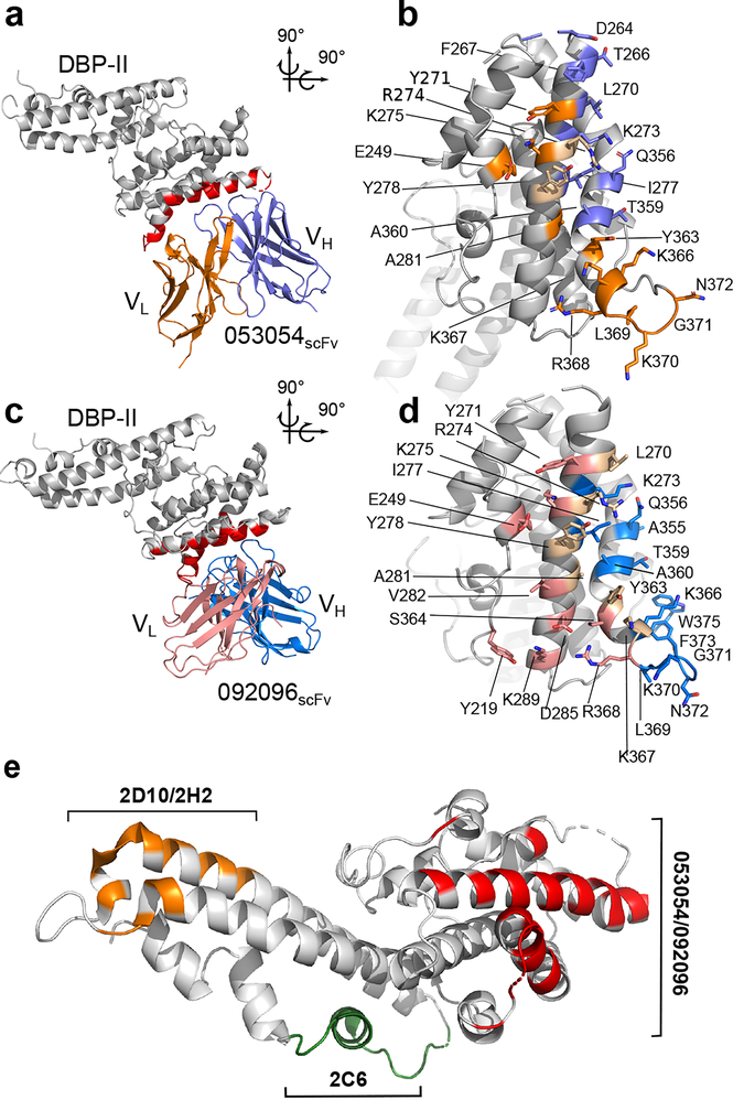
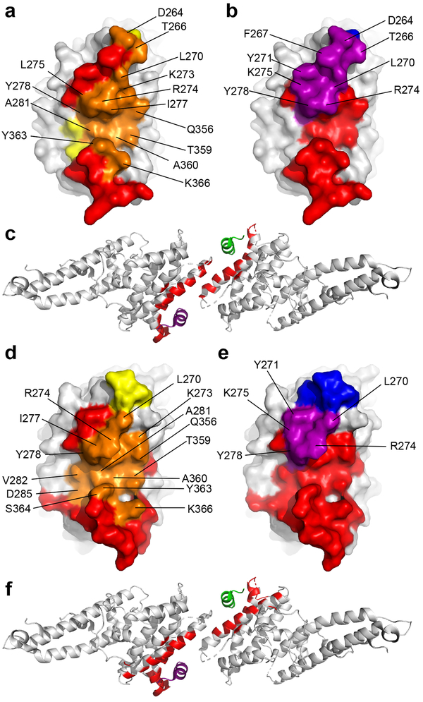
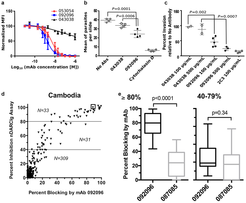
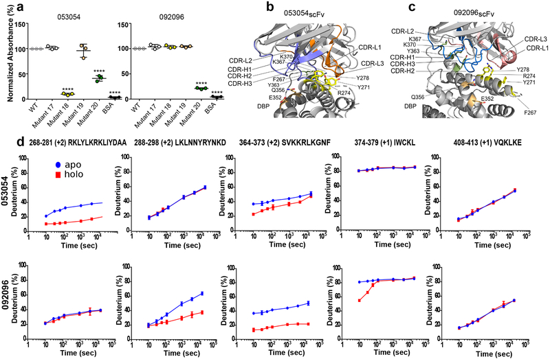
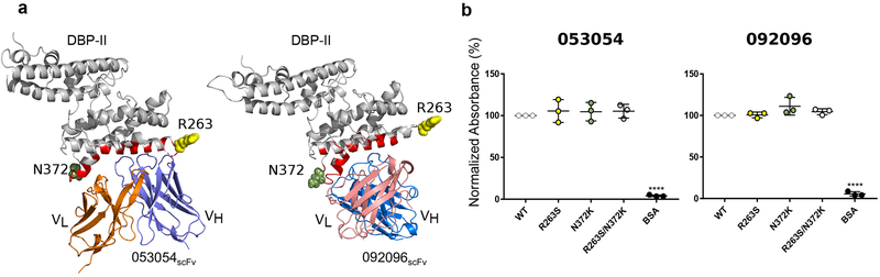
The Plasmodium vivax Duffy-binding protein (DBP) is a prime target of the protective immune response and a promising vaccine candidate for P. vivax malaria. Naturally acquired immunity (NAI) protects against malaria in adults residing in infection-endemic regions, and the passive transfer of malarial immunity confers protection. A vaccine that replicates NAI will effectively prevent disease. Here, we report the structures of DBP region II in complex with human-derived, neutralizing monoclonal antibodies obtained from an individual in a malaria-endemic area with NAI. We identified protective epitopes using X-ray crystallography, hydrogen-deuterium exchange mass spectrometry, mutational mapping and P. vivax invasion studies. These approaches reveal that naturally acquired human antibodies neutralize P. vivax by targeting the binding site for Duffy antigen receptor for chemokines (DARC) and the dimer interface of P. vivax DBP. Antibody binding is unaffected by polymorphisms in the vicinity of epitopes, suggesting that the antibodies have evolved to engage multiple polymorphic variants of DBP. The human antibody epitopes are broadly conserved and are distinct from previously defined epitopes for broadly conserved murine monoclonal antibodies. A library of globally conserved epitopes of neutralizing human antibodies offers possibilities for rational design of strain-transcending DBP-based vaccines and therapeutics against P. vivax.


Conflict of interest statement

COMPETING INTERESTS

The authors declare no competing interests.

Figures

Figure 1|
Figure 1|. Structural definition of human antibody epitopes in DBP.
a, Overall structure and epitope for the 053054 complex. Gray - DBP-II. Dark blue - 053054 heavy chain. Orange - 053054 light chain. Red - epitope. b, orthogonal detailed view of the epitope for 053054 in DBP. Gray - DBP-II. Dark blue – DBP residues contacted by the 053054 heavy chain. Orange – DBP residues contacted by the 053054 light chain. Beige – DBP residues contacted by both heavy and light chains. c, Overall structure and epitope for the 092096 complex. Gray - DBP-II. Light blue - 092096 heavy chain. Pink - 092096 light chain. Red - epitope. d, orthogonal detailed view of the epitope for 092096 in DBP. Gray - DBP-II. Light blue – DBP residues contacted by the 092096 heavy chain. Pink – DBP residues contacted by the 092096 light chain. Beige – DBP residues contacted by both heavy and light chains. e, Comparison of human and murine epitopes in DBP-II reveal epitopes are distinct. Orange - epitope of inhibitory murine mAbs 2D10/2H2. Green - epitope of inhibitory murine mAb 2C6. Red- epitopes of neutralizing human mAbs 053054 and 092096.
Figure 2|
Figure 2|. Neutralizing mAbs 053054 and 092096 block DARC-binding site and dimer interface.
a, Overlay of the 053054 epitope with the DARC-binding residues in DBP. Orange - overlap between the 053054 epitope and the DARC binding site. Yellow/Orange - DARC binding site. Red/Orange –the 053054 epitope. b, Overlay of 053054 epitope with the dimer interface of DBP. Purple - overlap between the 053054 epitope and the dimer interface. Blue/Purple - dimer interface. Red/Purple –the 053054 epitope. c, Mapping the 053054 epitope on the tetramer structure of DARC-bound DBP reveals the epitope overlaps with the DARC binding residues and dimer interface. Grey – DBP. Green – DARC monomer 1. Purple – DARC monomer 2. Red – epitope of 053054. d, Overlay of the 092096 epitope with the DARC-binding residues in DBP. Orange - overlap between the 092096 epitope and the DARC binding site. Yellow/Orange - DARC binding site. Red/Orange –the 092096 epitope. e, Overlay of 092096 epitope with the dimer interface of DBP. Purple - overlap between the 092096 epitope and the dimer interface. Blue/Purple - dimer interface. Red/Purple –the 092096 epitope. f, Mapping the 092096 epitope on the tetramer structure of DARC-bound DBP reveals the epitope overlaps with the DARC binding residues and dimer interface. Grey – DBP. Green – DARC monomer 1. Purple – DARC monomer 2. Red – epitope of 092096.
Figure 3|
Figure 3|. Human antibodies block DBP binding to RBCs and P. vivax invasion.
a, Inhibition of DBP-II binding to RBCs. The isotype matched mAb 043038 was used as a negative control. Data shown are mean ± SD of three biological replicates. b, Neutralization data for a Brazilian isolate of P.vivax invasion of human reticulocytes by human mAb 092096. Individual data points represent the total number of P. vivax infected cells per 20,000 RBCs and are shown as mean ± 1 SEM, for one P. vivax isolate performed in four biological replicate cultures in one experiment. Statistical differences between 092096 and no invasion inhibitor control (No Abs = medium alone) and non-inhibitory mAb 043038 were analyzed by one-way ANOVA and Dunnett’s test (p =0.0001 and 0.0006, respectively). Cytochalasin D, an actin inhibitor, served as a positive control for the inhibition of invasion. c, Neutralization data for Cambodian isolates of P.vivax invasion of human reticulocytes by human mAb 092096. Each data point represent results from one of five P. vivax isolates and the results presented as mean ± 1 SEM. These data represent results from five separate experiments, each with a different P. vivax isolate. All experiments had no Ab control and 4 out of 5 experiments used mouse anti-DARC monoclonal antibody 2C3 as a positive control and one experiment used heparin as positive control (11% invasion relative to no antibody, not shown in figure). The other controls were performed with only some experiments because of limited number of parasites available for different culture conditions. The isotype matched mAb 043038 serves as an additional negative control. Statistical comparison between non-inhibitory mAb 043038 (n=2) and neutralizling mAb 092096 was performed with one-way ANOVA and Dunnett’s test (p =0.002 for 100 μg/mL mAbs concentration, n=5, p =0.0007 for 500 μg/mL mAbs concentration, n=3). d, Human mAbs compete with epitopes recognized by serum from individuals with high levels of DBPII-specific blocking activity (≥80%) residing in Cambodia. Y-axis indicates the level of blocking activity using a DBPII:DARC binding assay, which measures the overall blocking activity in a serum sample. The x-axis is the percentage of blocking activity in the sample for which a given mAb competed. e, The box and whisker plots (median blocking activity, 25% and 75% interquartile ranges, and 95% confidence intervals) examine the degree to which mAbs 092096 and 087085 compete with serum blocking activity stratified as to whether participants had 80–100% blocking activity (n=33 for both mAbs, p<0.0001) versus lower activity of 40–79% (n=31 for both mAbs, p=0.34). The sample size for the two strata is shown in the scatter plots. Difference in blocking activity was examined using two-tailed Mann-Whitney U test.
Figure 4|
Figure 4|. Mutant ELISAs and HDX-MS reveal overlapping but distinct binding modes.
a, Evaluation of 053054 and 092096 binding to DBP Sal1 and four mutants by ELISA. Mutant 17: T257G, D258G, T259S, N260G, F261G, H262S. Mutant 18: F267A, Y271A, R274A, Y278A. Mutant 19: E352A, Q356A. Mutant 20: Y363A, K367A, K370A. Data are shown as mean ± 1 SD, calculated from three independent experiments with three technical replicates each. Statistical differences analyzed by one-way ANOVA and Dunnett’s test (****p < 0.0001). b, c, Position of DBP-II surface mutants tested in (a) with regards to the individual mAbs. The antibodies were structurally aligned in (b) and (c) (see also Supplementary Fig. 2) to show the different locations of the mutations with respect to each complex. DBP-II and antibody colored gray. Mutant 18 – yellow. Mutant 19 – salmon, Mutant 20 - green. d, HDX-MS kinetics for five DBP peptides in the presence (red) or absence (blue) of 053054 or 092096. Peptide sequence and charge state is shown (see also Supplementary Fig. 4 for full peptide coverage of DBP). The HDX data were determined in duplicate, and data are presented as mean +/− SD. For determinations where the deviation is not seen, it is smaller than the size of the data point. The statistical validity of the curves was also assured by making the measurement over seven time points. Given that the purpose of the HDX kinetic plots is to assign binding or no-binding, those assignments were made visually without further statistical analysis.
Figure 5|
Figure 5|. Strain-transcending human mAbs overcome polymorphisms located within the vicinity of their epitopes.
a, Location of polymorphisms R263S and N372K on DBP-II surface shown in DBP-II/053054 and DBP-II/092096 structures. Polymorphic residues 263 and 372 shown in ball representation in yellow and green, respectively. The location of R263 was modeled as this residue is located in a disordered segment that is not visible in either structure. b, ELISA for 053054 and 092096 against Sal-I DBP-II wild type and three DBP-II natural variants, R263S, N372K and double mutant R263S/N372K. Data are shown as mean ± SD, calculated from three independent experiments. Statistical differences between wild type Sal-1 DBP and the mutants were analyzed by one-way ANOVA and Dunnett’s test (****p < 0.0001).
Figure 6|
Figure 6|. Monoclonal antibody maturation.
Sequence identity analysis for 053054 and 092096 mAbs by a, heavy and b, light chain alignment. CDRs 1–3 are marked at the top of the alignment. Similar residues are highlighted gray, identical black. c-f, Amino acid sequence alignment of the variable heavy chain and light chain of the mAbs (c and d) 053054 and (e and f) 092096 against the germline V, D and J gene members with the highest sequence similarity defined by NCBI/IgBlast and IMGT/V-Quest indicated in each alignment. Complementarity determining regions (CDR 1–3) are shown at the top of the alignment. Amino acid mutations are highlighted green. Non-silent somatic hypermutations in CDRs are indicated by black stars. Red triangles mark insertions in the junction region. The secondary structure elements are indicated by blue arrows for β sheets and blue coils for α helices. g, The number of non-silent somatic mutations for CDRs of VH and VL chains of each antibody as well as junction insertions.

Comment in

References

    1. Guerra CA et al. The international limits and population at risk of Plasmodium vivax transmission in 2009. PLoS Negl Trop Dis 4, e774 (2010). - PMC - PubMed
    1. King CL et al. Naturally acquired Duffy-binding protein-specific binding inhibitory antibodies confer protection from blood-stage Plasmodium vivax infection. Proc Natl Acad Sci U S A 105, 8363–8 (2008). - PMC - PubMed
    1. Chootong P et al. Mapping epitopes of the Plasmodium vivax Duffy binding protein with naturally acquired inhibitory antibodies. Infect Immun 78, 1089–95 (2010). - PMC - PubMed
    1. Cole-Tobian JL et al. Age-acquired immunity to a Plasmodium vivax invasion ligand, the duffy binding protein. J Infect Dis 186, 531–9 (2002). - PubMed
    1. Grimberg BT et al. Plasmodium vivax invasion of human erythrocytes inhibited by antibodies directed against the Duffy binding protein. PLoS Med 4, e337 (2007). - PMC - PubMed

Publication types

MeSH terms