. 2019 Sep;4(9):1497-1507.
doi: 10.1038/s41564-019-0462-1. Epub 2019 May 27.

Structural basis for inhibition of Plasmodium vivax invasion by a broadly neutralizing vaccine-induced human antibody

Affiliations

Structural basis for inhibition of Plasmodium vivax invasion by a broadly neutralizing vaccine-induced human antibody

Thomas A Rawlinson et al. Nat Microbiol. 2019 Sep.

Abstract

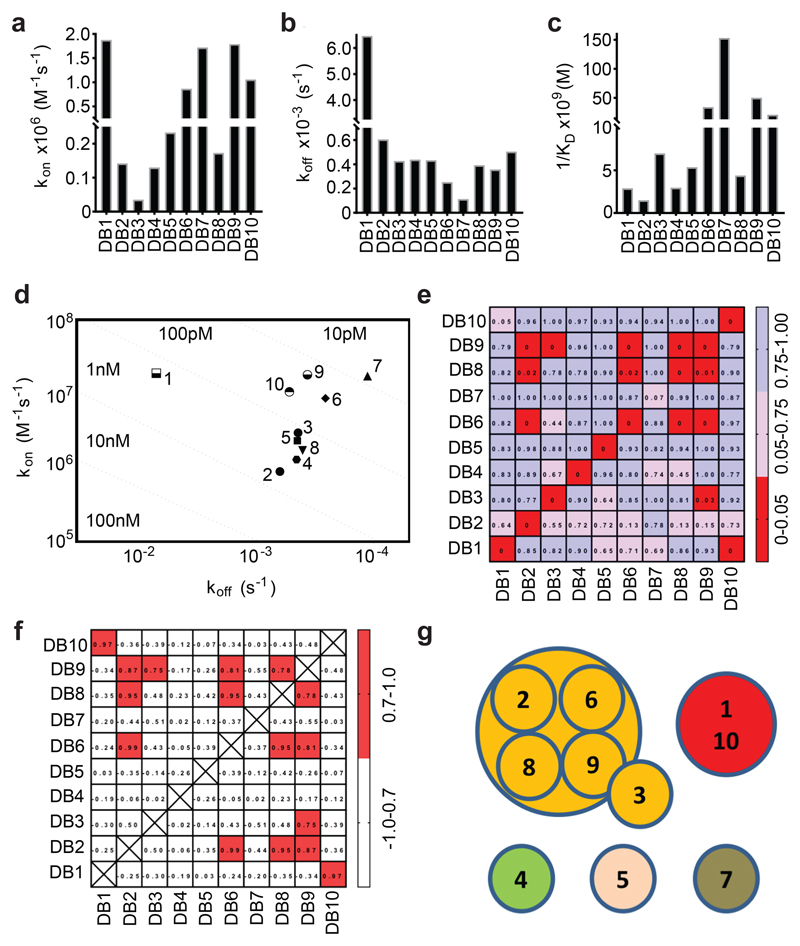
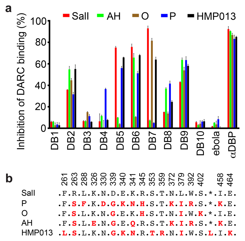
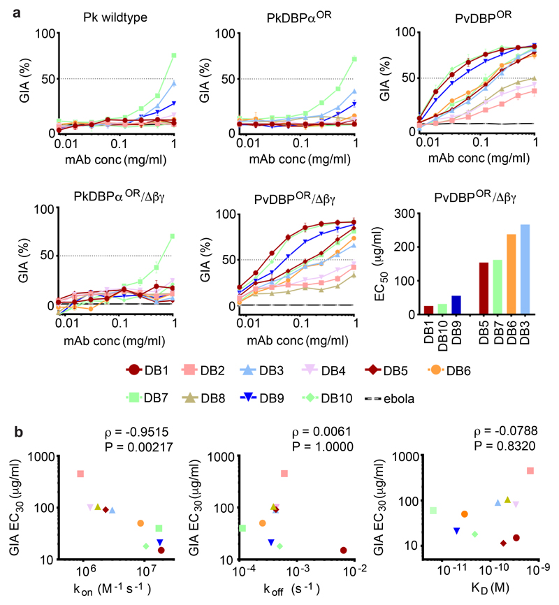
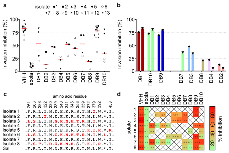
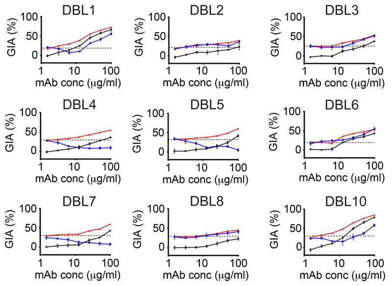
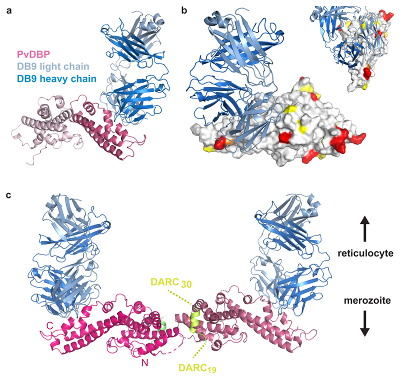
The most widespread form of malaria is caused by Plasmodium vivax. To replicate, this parasite must invade immature red blood cells through a process requiring interaction of the P. vivax Duffy binding protein (PvDBP) with its human receptor, the Duffy antigen receptor for chemokines. Naturally acquired antibodies that inhibit this interaction associate with clinical immunity, suggesting PvDBP as a leading candidate for inclusion in a vaccine to prevent malaria due to P. vivax. Here, we isolated a panel of monoclonal antibodies from human volunteers immunized in a clinical vaccine trial of PvDBP. We screened their ability to prevent PvDBP from binding to the Duffy antigen receptor for chemokines, and their capacity to block red blood cell invasion by a transgenic Plasmodium knowlesi parasite genetically modified to express PvDBP and to prevent reticulocyte invasion by multiple clinical isolates of P. vivax. This identified a broadly neutralizing human monoclonal antibody that inhibited invasion of all tested strains of P. vivax. Finally, we determined the structure of a complex of this antibody bound to PvDBP, indicating the molecular basis for inhibition. These findings will guide future vaccine design strategies and open up possibilities for testing the prophylactic use of such an antibody.


Conflict of interest statement

Competing Interests Statement

D.G.W.A, M.K.H. and S.J.D. are named inventors on patent applications relating to malaria vaccines, mAbs and/or immunization regimens.

Figures

Figure 1
Figure 1. Binding kinetics and epitope bins for the human anti-PvDBPII mAb panel.
Kinetic rate constants of binding for ten human mAbs (DB1-DB10) to PvDBPII as determined by SPR. (A) Association rate constant (kon); (B) Dissociation rate constant (koff); (C) Association constant (1/KD); and (D) Iso-affinity plot of kon against koff. (E) A “relative binding” matrix showing the fraction of a mAb bound to PvDBPII in the presence of a second bound mAb. Assays were conducted in both orientations. Boxes are colour-coded with ≥ 0.75 in blue, 0.75 > X > 0.05 in pink and ≤ 0.05 in red. Negative values were normalised to 0 and values >1 were normalised to 1. (F) A “binding profile correlation” matrix showing the Pearson product-moment correlation values of each mAb pair. The correlation threshold was set at 0.7; values equal to or above this are coloured in red as the threshold chosen to represent competition. (G) Epitope bins determined from E and F.
Figure 2
Figure 2. Inhibition of the binding of recombinant PvDBPII to DARC ectodomain.
(A) Assessment of the % binding of five naturally occurring variants of PvDBPII to the DARC ectodomain in vitro in the presence of 100 μg/mL concentration of each mAb (DB1-DB10). Individual titration curves are shown in Supplementary Figure 2. “αDBP” is polyclonal human anti-PvDBPII serum at 1:5 dilution while ‘ebola’ is an anti-Ebolavirus recombinant human IgG1 mAb included as a negative control. Data points represent the mean of three technical replicates, while the error bars represent the standard deviation. (B) Sequence polymorphisms of PvDBPII variants used in the assay. Numbering is according to the SalI reference sequence. Amino acid polymorphisms are indicated for the PvDBPII variants (P, O, AH and HMP013). Amino acids that are the same as the SalI reference sequence are black, those divergent from SalI are red and * indicates the absence of a leucine insertion between V429 and P430 in HMP013.
Figure 3
Figure 3. Growth inhibition of transgenic Plasmodium knowlesi (Pk) lines expressing PvDBP.
(A) Assays of growth Inhibitory activity (GIA) by the ten anti-PvDBPII human mAbs against five different Pk lines: ‘Wild type’ Pk (A1-H.1); PkDBPαOR; PkDBPαOR/Δβγ; PvDBPOR and PvDBPOR/Δβγ. Inhibition was tested in a two-fold dilution series starting at 1 mg/mL. Data points represent the mean of three technical replicates, while the error bars represent the standard deviation. The EC50 values (interpolated by non-linear regression) are shown in ascending rank order for the seven mAbs which reached >50 % GIA against PvDBPOR/Δβγ at the maximum concentration (1 mg/mL). (B) kon (on-rate), koff (off-rate) and KD (dissociation constant), plotted against GIA EC30 for PvDBPOR/Δβγ. EC30 values were used to include weaker-neutralising mAbs and were interpolated from non-linear regression curves. Kinetic data are as Figure 1 and Supplementary Table 2. Spearman’s rank correlation coefficient (ρ) and P value are shown.
Figure 4
Figure 4. Inhibition of invasion of reticulocytes by Thai Plasmodium vivax clinical isolates.
(A) Plasmodium vivax ex-vivo invasion assays were performed with thirteen separate isolates of infected blood from local patients. Each data point represents the % inhibition of each antibody against one of the thirteen isolates. All antibodies were tested at a final concentration of 1 mg/mL, except the positive control anti-DARC VHH (VHH) which was assayed at 25 μg/mL. The red bars represent the median % inhibition for each antibody. A recombinant human IgG1 anti-Ebolavirus mAb (ebola) was used as a negative control at 1mg/mL. (B) Percentage invasion inhibition by mAbs tested against the two Thai Plasmodium vivax isolates which share the PvDBPII gene sequence of SalI; isolate #4 (left hand bar of each pair) and isolate #7 (right hand bar of each pair). This demonstrates the degree of correlation with the hierarchy of mAb inhibition in the transgenic Plasmodium knowlesi GIA assays (Figure 3A). (C) Amino acid polymorphisms found within the PvDBPII gene segment of the eight Thai Pv isolates for which we obtained sequence information. The vaccine homologous SalI reference sequence is shown in the bottom row. Amino acids that are the same as the reference sequence black, divergent residues are red and * represents absence of a leucine insertion between V429 and P430 in the SalI reference sequence. (D) Summary matrix showing the percentage inhibition of invasion by each mAb for each of the sequenced strains. The positive control was the camelid anti-DARC nanobody (VHH) at 25 μg/mL and the negative control was a recombinant human IgG1 anti-Ebolavirus mAb (ebola) at a concentration of 1 mg/mL.
Figure 5
Figure 5. Assessment of synergy, additivity and antagonism by anti-PvDBPII human mAb combinations.
Assays of growth inhibitory activity (GIA) were performed to assess the inhibitory activity of DB9 in combination with the other mAbs against the PvDBPOR transgenic Plasmodium knowlesi line. DB9 was held at a fixed concentration of 25 μg/mL, while the other mAbs are in a two-fold dilution series starting at 100 μg/mL. The black line shows the % inhibition of each mAb in the absence of DB9. The red line shows the predicted additive inhibition (‘Bliss additivity’ as calculated in the equation in Methods) of the indicated mAb plus DB9 at 25 μg/mL. The blue line shows the actual observed % inhibition of the two combined mAbs. The dotted black line gives the % inhibition of DB9 alone at 25 μg/mL. Data points represent the mean of three technical replicates, while the error bars represent the standard deviation.
Figure 6
Figure 6. The structural basis for inhibition of PvDBPII by antibody DB9.
(A) The structure of PvDBPII (pink) bound to the Fab fragment of DB9 (blue). PvDBPII is shown in two shades of pink, with subdomains 1 and 2 in light pink and subdomain 3 in dark pink. DB9 is in two shades with the light chain in light blue and the heavy chain in dark blue. (B) PvDBPII is shown in surface representation in grey, with residues known to be polymorphic highlighted according to their sequence entropy (yellow = 0.15-0.3, orange = 0.3-0.45 and red > 0.45). DB9 is in blue, and binds to a conserved region of PvDBPII. (C) The structure of PvDBPII:DB9 complex superimposed on the structure of the PvDBPII dimer structure bound to a peptide from the DARC ectodomain, indicating that DB9 may prevent the binding of PvDBP to DARC in the context of the reticulocyte membrane.

Comment in

References

    1. WHO. Control and elimination of plasmodium vivax malaria: a technical brief. 2015. ISBN 978 92 4 150924 4.
    1. Gething PW, et al. A long neglected world malaria map: Plasmodium vivax endemicity in 2010. PLoS Negl Trop Dis. 2012;6:e1814. - PMC - PubMed
    1. Gruszczyk J, et al. Transferrin receptor 1 is a reticulocyte-specific receptor for Plasmodium vivax. Science. 2018;359:48–55. - PMC - PubMed
    1. Malleret B, Renia L, Russell B. The unhealthy attraction of Plasmodium vivax to reticulocytes expressing transferrin receptor 1 (CD71) Int J Parasitol. 2017;47:379–383. - PubMed
    1. Singh SK, Hora R, Belrhali H, Chitnis CE, Sharma A. Structural basis for Duffy recognition by the malaria parasite Duffy-binding-like domain. Nature. 2006;439:741–4. - PubMed

Publication types

MeSH terms