Review
. 2019 Dec;12(6):626-636.
doi: 10.1007/s12328-019-00996-6. Epub 2019 May 27.

Isolated pancreatic metastasis from malignant melanoma: a case report and literature review

Affiliations
Review

Isolated pancreatic metastasis from malignant melanoma: a case report and literature review

Yoshifumi Nakamura et al. Clin J Gastroenterol. 2019 Dec.

Abstract

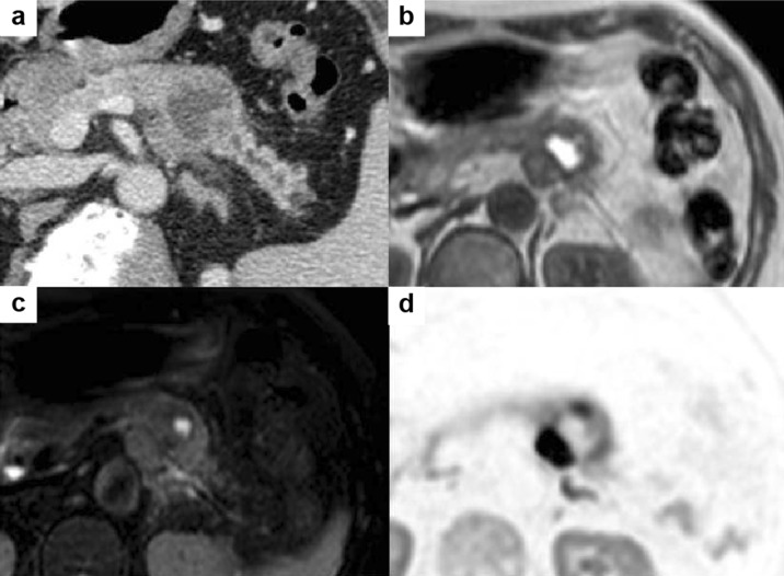
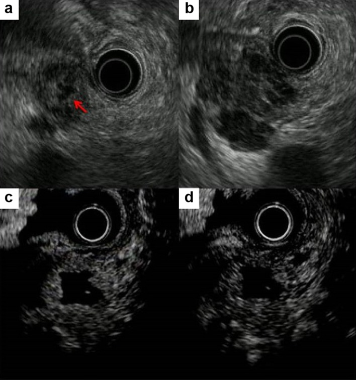
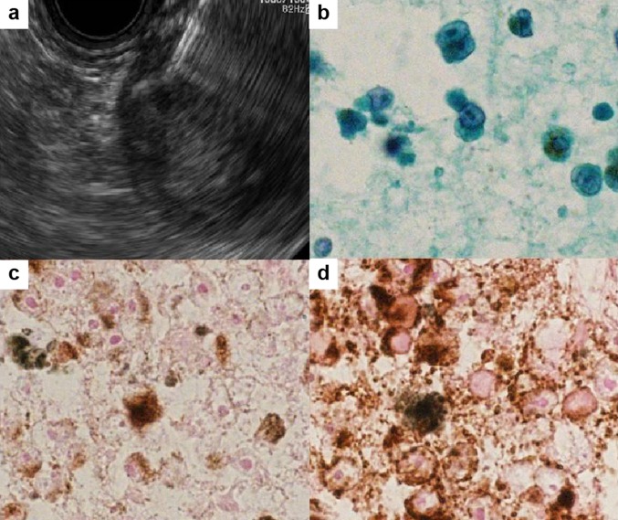
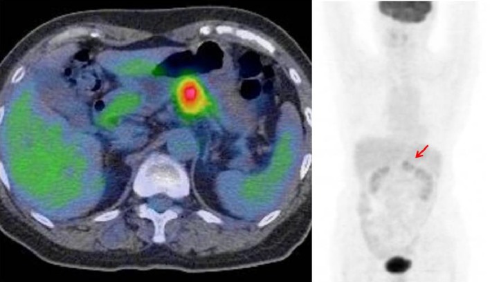
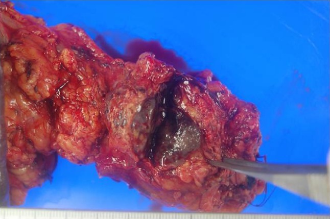
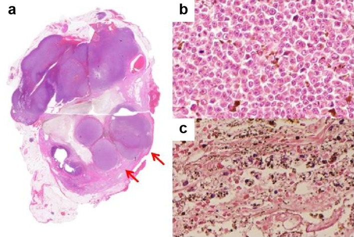
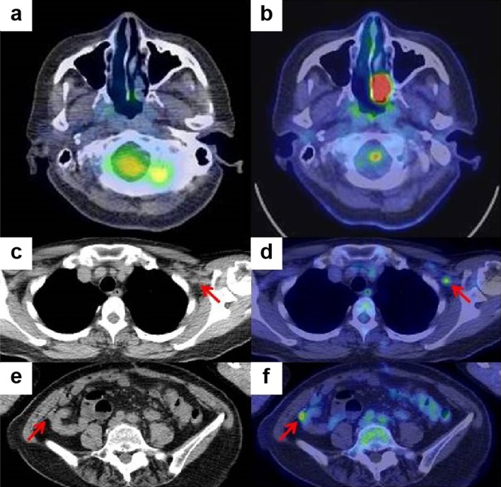
Isolated pancreatic metastasis from malignant melanoma is rare. Pancreatic metastasis is difficult to diagnose in patients with unknown primary malignant melanoma. Endoscopic ultrasound-guided fine-needle aspiration plays an important role in confirming the diagnosis. A 67-year-old woman was referred to our institution because of a mass in her pancreas. Computed tomography and magnetic resonance imaging revealed a 35-mm mass localized on the pancreatic tail, with low attenuation, surrounded by a high-attenuation rim. Endoscopic ultrasonography revealed a hypoechoic mass with central anechoic areas. Endoscopic ultrasound-guided fine-needle aspiration of the mass was performed, and the pathological diagnosis was malignant melanoma. Intense fluorodeoxyglucose uptake was observed in the pancreatic tail on positron emission tomography-computed tomography. No other malignant melanoma was found. Distal pancreatectomy was performed. Six months postoperatively, positron emission tomography-computed tomography revealed high uptake in the left nasal cavity, and biopsy revealed the mass to be a malignant melanoma, indicating that the primary site of the malignant melanoma was the left nasal cavity and that the pancreatic mass and peritoneal lesion were metastases. The patient had survived > 2 years after the distal pancreatectomy. Pancreatic resection of isolated pancreatic metastasis can possibly prolong survival; however, metastatic melanoma usually has poor prognosis.

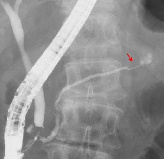
Keywords: Case report; Endoscopic retrograde cholangiopancreatogram (ERCP); Endoscopic ultrasound (EUS); Endoscopic ultrasound-guided fine-needle aspiration (EUS-FNA); Malignant melanoma.


Conflict of interest statement

The authors declare no conflicts of interest or financial arrangement with any company.

Figures

Fig. 1
Fig. 1
Computed tomography image. a Mass in the tail of the pancreas with pancreatic ductal dilation. The central mass is hyperintense on T1-weighted image (b) and hypointense on T2-weighted image (c). d Peripheral rim of the mass is hyperintense on diffusion-weighted image
Fig. 2
Fig. 2
Endoscopic retrograde cholangiopancreatogram revealed smooth narrowing and displacement of the pancreatic duct with upstream dilatation
Fig. 3
Fig. 3
Endoscopic ultrasonography revealed hypoechoic and homogenous heterogeneous mass (a) with central anechoic areas (b, arrow). Contrast-enhanced endoscopic ultrasonography shows isoenhancement at 20 s (c) and hypoenhancement at 120 s (d) with central non-enhancement of the peripheral rim of the mass
Fig. 4
Fig. 4
a Endoscopic ultrasound-guided fine-needle aspiration of the peripheral rim of the mass. b Cytologic results revealed a large nucleus and a high nuclear/cytoplasmic ratio in the cells, with brown pigmentation. Immunocytochemical staining with Melan A (c) and Human Melanoma Black 45 (d)
Fig. 5
Fig. 5
Intense fluorodeoxyglucose uptake only in the body and tail of the pancreas (arrow)
Fig. 6
Fig. 6
Resected surgical specimen showing a black–brown mass in the tail of the pancreas
Fig. 7
Fig. 7
a Loupe image of the resection specimen. The peripheral rim of the mass has nodular components (arrows). b Tumor cells in the peripheral rim of the mass have anisokaryosis and clear nuclei with melanin production. c Center of the mass was necrotic
Fig. 8
Fig. 8
Positron emission tomography–computed tomography image of the nasal cavity before (a) and after surgery (b). Plane computed tomography and positron emission tomography–computed tomography images after surgery revealed left infraclavicular lymph node metastasis (c, d) and a small peritoneal nodule (e, f)

Similar articles

Cited by

References

    1. Jana T, Caraway NP, Irisawa A, et al. Multiple pancreatic metastases from malignant melanoma: conclusive diagnosis with endoscopic ultrasound-guided fine needle aspiration. Endosc Ultrasound. 2015;4:145–148. - PMC - PubMed
    1. Larsen AK, Krag C, Geertsen P, et al. Isolated malignant melanoma metastasis to the pancreas. Plast Reconstr Surg Glob Open. 2013;1:e74. - PMC - PubMed
    1. De Moura DT, Chacon DA, Tanigawa R, et al. Pancreatic metastases from ocular malignant melanoma: the use of endoscopic ultrasound-guided fine-needle aspiration to establish a definitive cytologic diagnosis: a case report. J Med Case Rep. 2016;10:332. - PMC - PubMed
    1. Portale TR, Di Benedetto V, Mosca F, et al. Isolated pancreatic metastasis from melanoma. Case report. G Chir. 2011;32:135–137. - PubMed
    1. Birnbaum DJ, Moutardier V, Turrini O, et al. Isolated pancreatic metastasis from malignant melanoma: is pancreatectomy worthwhile? J Surg Tech Case Rep. 2013;5:82–84. - PMC - PubMed

MeSH terms