Review
. 2019 Dec;56(12):7950-7965.
doi: 10.1007/s12035-019-1641-z. Epub 2019 May 27.

Molecular Plasticity of the Nucleus Accumbens Revisited-Astrocytic Waves Shall Rise

Affiliations
Review

Molecular Plasticity of the Nucleus Accumbens Revisited-Astrocytic Waves Shall Rise

Julianna Kardos et al. Mol Neurobiol. 2019 Dec.

Abstract

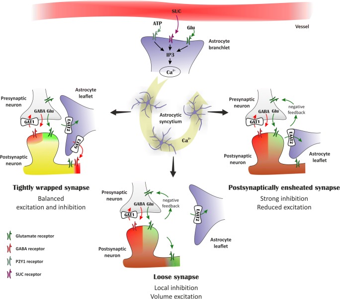
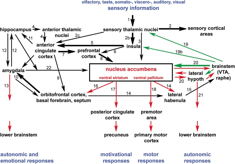
Part of the ventral striatal division, the nucleus accumbens (NAc) drives the circuit activity of an entire macrosystem about reward like a "flagship," signaling and leading diverse conducts. Accordingly, NAc neurons feature complex inhibitory phenotypes that assemble to process circuit inputs and generate outputs by exploiting specific arrays of opposite and/or parallel neurotransmitters, neuromodulatory peptides. The resulting complex combinations enable versatile yet specific forms of accumbal circuit plasticity, including maladaptive behaviors. Although reward signaling and behavior are elaborately linked to neuronal circuit activities, it is plausible to propose whether these neuronal ensembles and synaptic islands can be directly controlled by astrocytes, a powerful modulator of neuronal activity. Pioneering studies showed that astrocytes in the NAc sense citrate cycle metabolites and/or ATP and may induce recurrent activation. We argue that the astrocytic calcium, GABA, and Glu signaling and altered sodium and chloride dynamics fundamentally shape metaplasticity by providing active regulatory roles in the synapse- and network-level flexibility of the NAc.

Keywords: Astrocytic endfeets; Mixed GABAergic and Gluergic synapses; Motivation-reward metaplasticity; Nucleus accumbens macrosystem; Perisynaptic astrocytic processes; Succinate receptor.


Conflict of interest statement

The authors declare that they have no competing interests.

Figures

Fig. 1
Fig. 1
Astrocytes may dynamically control plasticity of mixed inhibitory and excitatory synapses. In the tightly wrapped synapse (left), Glu transporters facing the synapse can quickly take up released Glu, thereby preventing the activation of extrasynaptic Glu receptors. The simultaneous activation of synaptic GABA and Glu receptors results in balanced excitation and inhibition. During intense excitation, EAAT2 activity may also induce GABA release by reverse transport, thereby generating tonic inhibition [68, 69]. When astrocytic leaflets are withdrawn from the synapse (bottom), reduced Glu uptake leads to activation of presynaptic mGluRs inhibiting further Glu release and of extrasynaptic Glu receptors increasing tonic excitation. Asymmetric astrocytic coverage of axonal boutons and dendritic spines (right) [51] favors reduced Glu release by activating presynaptic mGluRs, resulting in a dominantly inhibitory response following GABA/Glu co-release
Fig. 2
Fig. 2
Afferent and efferent connections of the accumbens/ventral striatum. The scheme represents the neuronal pathway interconnectivity converging onto and originating from the nucleus accumbens (NAc)/ventral striatum/pallidum. Different modalities of sensory information reach the NAc through the sensory thalamic nuclei directly and indirectly, too. These glutamatergic inputs (black arrows) are combined in the NAc with monoaminergic (dopaminergic and serotonergic) inputs (green) from the ventral tegmental area (VTA) and the raphe nuclei, respectively. The projections from the NAc are inhibitory (red arrows). Efferent projections to cerebral brain regions initiate motivational and motor responses, while indirect output to the lateral hypothalamus and the amygdala initiates autonomic and emotional responses. Thick arrows represent particularly massive projections. The white matter tracts where the different pathways are located are indicated by numbers as follows: (1) ascending somato- and viscerosensory pathways to the thalamus; (2) thalamocortical radiation; (2b) sensory inputs to the posterior insular cortex; (2c) sensory inputs to the anterior cingulate cortex; (3) thalamo-striatal and pallidal projections; (4) hippocampal-anterior thalamic-anterior cingulate cortex connections through the “Papez circle”; (5) anterior insular projections to the anterior cingulate cortex (the two hubs of the salience network); (6) direct anterior insular projections to the prefrontal cortex (to the ventrolateral and dorsolateral prefrontal cortex); (7) bilateral anterior cingular-prefrontal cortical fiber connections; (8) prefrontal neuronal feedback to the nucleus accumbens; (9) bilateral anterior cingular-orbitofrontal cortical fiber connections; (10) bilateral connections between the orbitofrontal cortex and the amygdala (uncinate fascicle); (11) anterior cingulate projections to the hippocampus through the parahippocampal cortex; (12) amygdala connections with the hippocampus (via peri- and entorhinal cortex); (13) descending amygdala projections to the lower brainstem (partly through the stria terminalis); (14) stria medullaris thalami; (15) fasciculus retroflexus; (16) nucleus accumbens, ventral striatal/pallidal projections to the orbitofrontal cortex, basal forebrain, and septum; (17) fibers from the nucleus accumbens/ventral striatal and pallidal neurons in the fronto-parietal neuronal connections (“dorsal default mode network”); (18) nucleus accumbens, ventral striatal/pallidal projections to the premotor and motor cortical areas; (19) ascending brainstem dopaminergic (from the ventral tegmental area) and serotinergic fibers (from the midline midbrain raphe nuclei) to the thalamus (one portion of the ascending reticular activating system); (19b) ascending brainstem dopaminergic and serotinergic fibers to the insula; (20) medial forebrain bundle; (21) descending fibers from the lateral hypothalamus to the lower brainstem; (22) ventral amygdalofugal pathway

References

    1. Abraham WC, Bear MF. Metaplasticity: the plasticity of synaptic plasticity. Trends Neurosci. 1996;19:126–130. doi: 10.1016/S0166-2236(96)80018-X. - DOI - PubMed
    1. Abraham WC. Metaplasticity: tuning synapses and networks for plasticity. Nat Rev Neurosci. 2008;9:387–399. doi: 10.1038/nrn2356. - DOI - PubMed
    1. Adamsky A, Kol A, Kreisel T, Doron A, Ozeri-Engelhard N, Melcer T, Rafaeli R, Horn H, Regev L, Groysman M, London M, Goshen I. Astrocytic activation generates de novo neuronal potentiation and memory enhancement. Cell. 2018;174:59–71. doi: 10.1016/j.cell.2018.05.002. - DOI - PubMed
    1. Allen NJ, Eroglu C. Cell biology of astrocyte-synapse interactions. Neuron. 2017;96:697–708. doi: 10.1016/j.neuron.2017.09.056. - DOI - PMC - PubMed
    1. Al-Muhtasib N, Forcelli PA, Vicini S. Differential electrophysiological properties of D1 and D2 spiny projection neurons in the mouse nucleus accumbens core. Physiol Rep. 2018;6:e13784. doi: 10.14814/phy2.13784. - DOI - PMC - PubMed

LinkOut - more resources