Review
. 2019 Nov 26;58(47):4657-4666.
doi: 10.1021/acs.biochem.9b00260. Epub 2019 Jun 11.

Mechanochemistry in Translation

Affiliations
Review

Mechanochemistry in Translation

Sarah E Leininger et al. Biochemistry. .

Abstract

As the influence of translation rates on protein folding and function has come to light, the mechanisms by which translation speed is modulated have become an important issue. One mechanism entails the generation of force by the nascent protein. Cotranslational processes, such as nascent protein folding, the emergence of unfolded nascent chain segments from the ribosome's exit tunnel, and insertion of the nascent chain into or translocation of the nascent chain through membranes, can generate forces that are transmitted back to the peptidyl transferase center and affect translation rates. In this Perspective, we examine the processes that generate these forces, the mechanisms of transmission along the ribosomal exit tunnel to the peptidyl transferase center, and the effects of force on the ribosome's catalytic cycle. We also discuss the physical models that have been developed to predict and explain force generation for individual processes and speculate about other processes that may generate forces that have yet to be tested.


Conflict of interest statement

The authors declare no competing financial interest.

Figures
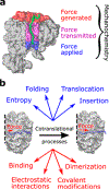
Figure 1
Figure 1
Cotranslational processes generate force that acts on the catalytic core of the ribosome. (a) Cotranslational processes involve the nascent chain (green) and occur outside or at the cytoplasmic end of the ribosomal exit tunnel (red region), and the forces they generate can be transmitted through the nascent chain inside the exit tunnel (purple region) to the PTC (located in the blue region), where they can act on peptide bond formation and potentially other substeps of the ribosome’s catalytic cycle. (b) Four cotranslational processes have been shown to generate forces (blue). Many other cotranslational processes and interactions have the potential to generate forces because they cause changes in free energy but have yet to be tested (red).
Figure 2
Figure 2
Unstructured nascent chain segments can generate a pulling force. The nascent chains in panels a and b are the same length; however, the N-terminus in panel a remains within the exit tunnel where it is more confined, while the N-terminus in panel b has left the exit tunnel and is free to adopt a wide variety of conformations. This increases the entropy of the system and decreases the free energy of the system, generating an entropic pulling force transmitted back to the PTC.
Figure 3
Figure 3
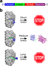
Cotranslational protein folding generates forces. (a) Arrest sequence assays use protein constructs similar to that shown. They start with a domain of interest at the N-terminus, attach a linker of variable length, attach an arrest sequence, and occasionally have a reporter domain such as GFP at the C-terminus. (b) Experiments and simulations have shown that there is a narrow range of linker lengths where folding forces occur. Simulations have shown in molecular detail that short linkers (top) do not allow the domain to cotranslationally fold, so no force is generated (and arrest sequence assays exhibit a correspondingly small fraction of full-length protein produced). Long linkers (bottom) allow the domain to fold; however, the domain has fewer contacts with the ribosome that are needed to generate force, and there is more slack in the linker, which weakens its ability to transmit force if any is generated. Linkers of an ideal length (middle) allow the domain to fold and push against the ribosome, with little slack in the linker, making force generation and transmission as efficient as possible. It is at these lengths where higher fractions of full-length protein are observed in arrest sequence assays.
Figure 4
Figure 4
Experimental and simulation results are in good agreement for cotranslational folding and insertion. (a) Simulation and experimental results agree well for the cotranslational folding of a Titin I27 domain. Panel a modified from ref . (b) Simulation and (c) experimental results show similarly aligned force peaks for the cotranslational insertion of polyleucine hydrophobic segments into a membrane. Blue and green dotted lines added for emphasis. Panels b and c modified from ref .
Figure 5
Figure 5
Statistical mechanical model (eq 1) that can accurately describe the results from varied data sets. Equation 1 was developed to describe simulation data (blue) of protein 1F0Z and utilizes the free energies of the folded protein on the ribosome at a given linker length. The experimental arrest sequence data (red) report the fraction of full-length protein produced vs free energies of mutated S6 protein in the absence of the ribosome. However, the ribosome is known to destabilize folded domains. To account for this, we added a constant value of 5.75 kcal/mol to all of the experimentally reported stabilities, which shifts the free energy of the domains such that reported half-maximum fraction full length is at ΔGUN,L = 0. We find the model fFL=fFL,NfFL,U1+eβ(ΔGUN,L+5.75) [where fFL is the fraction of full-length protein produced and fFL,N and fFL,U are the characteristic fractions of full-length protein produced if the domain was in the folded (N) and unfolded (U) states, respectively] describes the trend in the experimental data well. Note that the left y-axis corresponds to the force for the data presented as blue circles while the right y-axis corresponds to the fraction full-length data presented as red triangles. Data for S6 taken from ref . Data for protein 1F0Z and the model taken from ref .
Figure 6
Figure 6
Topology and translation speed modulate the forces generated during cotranslational folding. (a) Linear representations of wild-type and rewired protein 1P9K, with helices shown as cylinders and strands represented by arrows. (b) Crystal structures of these domains, colored from red (N-terminus) to blue (C-terminus). (c) The presence of C-terminal β-hairpins leads to the generation of large forces. (d) In the case of continuous synthesis, folding of the 2IST domain is delayed by six residues. (e) When the translation rate is faster than the folding rate (purple triangles and red squares), folding domains (data for 2IST with a C-terminal linker of variable length) generate less force because they fold at longer linker lengths. Figure modified from ref .
Figure 7
Figure 7
Examples of domains (a) with and (b) without C-terminal hairpins. For this analysis, we defined C-terminal hairpin structures as the last two secondary structural elements in a domain, each with at least four residues, which are antiparallel and connected by a loop and form at least three van der Waals contacts. Hairpin-containing structures are PDB entries 1QYS, 3GB1, 1A6J, and 1LOU (from left to right, respectively). Structures lacking hairpins are 1BIA, 1NY8, 2JSO, and 1DXE (from left to right, respectively).
Figure 8
Figure 8
Nascent protein backbone that is the primary route of force transmission through the exit tunnel. Fritch and co-workers used a series of simulations to show that force is transmitted through the protein backbone (black arrow) and not the tunnel walls (yellow arrow).
Figure 9
Figure 9
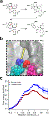
Tensile forces reduce the free energy barrier to peptide bond formation. (a) Structural representation of the reaction corresponding to peptide bond formation, with the movement of electrons colored red. (b) Force vector acting on the P-site residue. The ribosome (gray) is cropped to show only the portions near the PTC. The P-site residue is colored blue (the other nascent chain residues have been omitted for the sake of clarity), and the P-site tRNA is colored cyan. When in an extended conformation, the nascent chain pulls at an angle 15° below the long axis of the exit tunnel (the yellow arrow shows the direction of the force vector reported in ref 22), which reduces the free energy barrier to peptide bond formation between the P-site residue and the A-site residue (red), which is attached to its tRNA (magenta). (c) Quantum mechanical/molecular mechanical calculations show that when tensile forces are present (in this case due to the entropic pulling force arising from a longer unstructured nascent chain as compared to a shorter one), a lower free energy barrier to peptide bond formation occurs. Panels a and c modified from ref .

References

    1. Takacs L (2013) The historical development of mechano-chemistry. Chem. Soc. Rev 42, 7649–7659. - PubMed
    1. Howard JL, Cao Q, and Browne DL (2018) Mechanochemistry as an emerging tool for molecular synthesis: what can it offer? Chem. Sci 9, 3080–3094. - PMC - PubMed
    1. Do JL, and Friščić T (2017) Mechanochemistry: A Force of Synthesis. ACS Cent. Sci 3, 13–19. - PMC - PubMed
    1. Wang GW, Komatsu K, Murata Y, and Shiro M (1997) Synthesis and X-ray structure of dumb-bell-shaped C120. Nature 387, 583–586.
    1. Komatsu K, Wang GW, Murata Y, Tanaka T, Fujiwara K, Yamamoto K, and Saunders M (1998) Mechanochemical Synthesis and Characterization of the Fullerene Dimer C120. J. Org. Chem 63, 9358–9366.

Publication types

Substances