. 2019 Jun 1;142(6):1561-1572.
doi: 10.1093/brain/awz102.

FAHN/SPG35: a narrow phenotypic spectrum across disease classifications

Affiliations

FAHN/SPG35: a narrow phenotypic spectrum across disease classifications

Tim W Rattay et al. Brain. .

Abstract

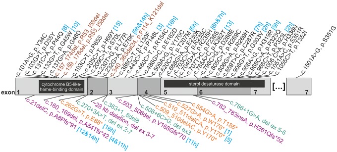
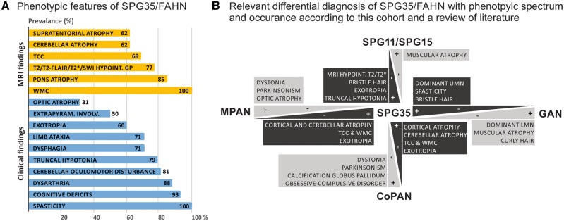
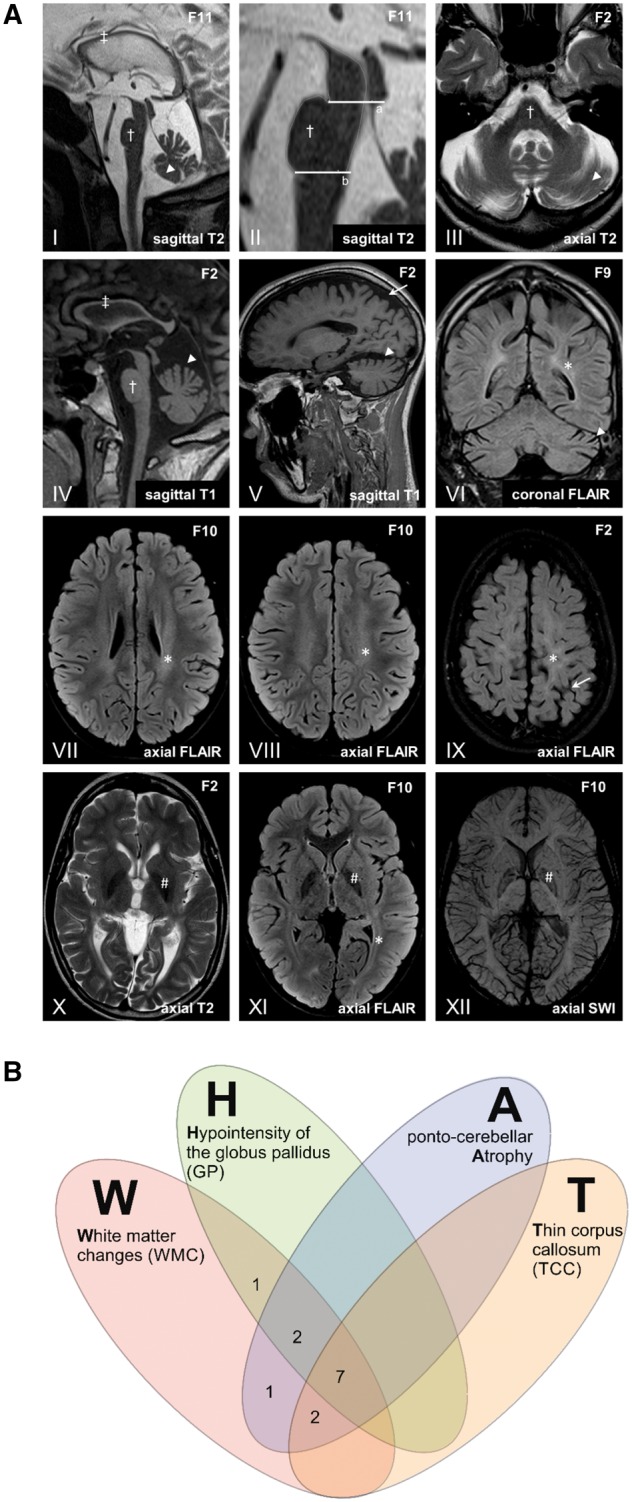
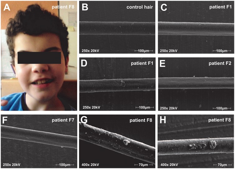
The endoplasmic reticulum enzyme fatty acid 2-hydroxylase (FA2H) plays a major role in the formation of 2-hydroxy glycosphingolipids, main components of myelin. FA2H deficiency in mice leads to severe central demyelination and axon loss. In humans it has been associated with phenotypes from the neurodegeneration with brain iron accumulation (fatty acid hydroxylase-associated neurodegeneration, FAHN), hereditary spastic paraplegia (HSP type SPG35) and leukodystrophy (leukodystrophy with spasticity and dystonia) spectrum. We performed an in-depth clinical and retrospective neurophysiological and imaging study in a cohort of 19 cases with biallelic FA2H mutations. FAHN/SPG35 manifests with early childhood onset predominantly lower limb spastic tetraparesis and truncal instability, dysarthria, dysphagia, cerebellar ataxia, and cognitive deficits, often accompanied by exotropia and movement disorders. The disease is rapidly progressive with loss of ambulation after a median of 7 years after disease onset and demonstrates little interindividual variability. The hair of FAHN/SPG35 patients shows a bristle-like appearance; scanning electron microscopy of patient hair shafts reveals deformities (longitudinal grooves) as well as plaque-like adhesions to the hair, likely caused by an abnormal sebum composition also described in a mouse model of FA2H deficiency. Characteristic imaging features of FAHN/SPG35 can be summarized by the 'WHAT' acronym: white matter changes, hypointensity of the globus pallidus, ponto-cerebellar atrophy, and thin corpus callosum. At least three of four imaging features are present in 85% of FA2H mutation carriers. Here, we report the first systematic, large cohort study in FAHN/SPG35 and determine the phenotypic spectrum, define the disease course and identify clinical and imaging biomarkers.

Keywords: FA2H; FAHN; SPG35; hereditary spastic paraplegia; imaging biomarker.


Figures

Figure 1
Figure 1
Mutation spectrum in FA2H/SPG35. Structure of the FA2H coding sequence (NCBI Reference Sequence: NM_024306.4) is shown with numbered exons 1–7 in alternating grey shades. For each variant, cDNA change and the presumed effect on the resulting protein are given (where possible). For changes observed in this study, the family number is given in square brackets and turquoise font; the letter ‘h’ indicates (compound)-heterozygous occurrence. Missense mutations (black) and inframe deletions (brown) are listed above, truncating mutations including nonsense mutations (orange), frameshift insertions or deletions (purple) and splice changes (green) below the graph. The cDNA is overlain by the two known functional protein domains (cytochrome B5-like heme-binding domain and sterol desaturase domain), indicated as black boxes.
Figure 2
Figure 2
Phenotypic spectrum of FAHN/SPG35 and relevant differential diagnosis. (A) The phenotypic spectrum of FAHN/SPG35 with prevalence rates (in per cent) of specific features. MRI findings are coloured in yellow, clinical signs and symptoms are highlighted in blue. (B) The FAHN/SPG35 phenotype overlaps the clinical presentation of a range of movement disorders. Main differential diagnoses of FAHN/SPG35 from the spectrum of hereditary spastic paraplegias (cHSPs), NBIA and giant axonal neuropathy that shares some features of the ultrastructural hair phenotypes with FAHN/SPG35 are depicted. Discriminating features are highlighted. CoPAN = COASY protein-associated neurodegeneration; GAN = giant axonal neuropathy; MPAN = mitochondrial membrane protein-associated neurodegeneration.
Figure 3
Figure 3
MRI findings in FAHN/SPG35. (A) The most frequent imaging findings in FAHN/SPG35 can be summarized by the acronym ‘WHAT’ (i) white matter changes (asterisk in VI–IX and XI); (ii) hypointensity of the globus pallidus in T2/T2-FLAIR/T2*/SWI (hash symbol in X–XII); (iii) ponto-cerebellar atrophy (dagger and arrowhead in I–VI); and (iv) thin corpus callosum. Sagittal images show a general corpus callosum thinning predominantly affecting the (dorsal) body (double dagger in I and IV). Pontine atrophy (dagger symbol in I–IV) was measured on midsagittal MRI (II) with the upper border a line (‘a’) through the superior pontine notch and the inferior edge of the quadrigeminal plate, and with the lower border (‘b’) parallel to the upper border and through the inferior pontine notch. Ponto (dagger)- cerebellar (arrowhead) atrophy is depicted in III with widening of the fourth ventricle, affecting the upper vermis and the hemispheres (I, III-VI). Predominantly mild supratentorial atrophy with a focus on the parietal lobe is depicted in V and IX (arrows). Periventricular white matter abnormalities are shown on T2-FLAIR images most affecting the parietal white matter (asterisk in VI–IX and XII). Hypointense signal of the globus pallidus associated with iron accumulation is shown on T2, T2-FLAIR and susceptibility weighted images (hash symbol in X–XII). (B) Venn diagram depicting the occurrence of MRI features contributing to the ‘WHAT’ imaging phenotype. In 85% of cases (11/13), at least three of four ‘WHAT’ features were found, in 54% (7/13), all four features were present.
Figure 4
Figure 4
Hair analysis: macroscopy and electron microscopic finding. (A) Photograph of Patient F8 with bristle like hair structure, which is kept short by the caregivers. (BH) Scanning electron microscopy images at 250× (BF) and 400× (G and H) of representative hair shafts. (B) Healthy control hair. (C and D) Patient F1 exemplary hair shafts present with longitudinal grooves in C and adhesive plaques in D. (E) Hair of Patient F2 shows longitudinal grooves but no adhesive plaques. (F) Longitudinal grooves in hair of Patient F7. (G and H) Hair of Patient F8 shows longitudinal grooves and adhesive plaques with a chunky appearance.

References

    1. Astudillo L, Sabourdy F, Therville N, Bode H, Segui B, Andrieu-Abadie N et al. . Human genetic disorders of sphingolipid biosynthesis. J Inherit Metab Dis 2015; 38: 65–76. - PubMed
    1. Darley FL, Aronson AE, Brown JR. Differential diagnostic patterns of dysarthria. J Speech Hear Res 1969; 12: 246–69. - PubMed
    1. Dick KJ, Eckhardt M, Paisan-Ruiz C, Alshehhi AA, Proukakis C, Sibtain NA et al. . Mutation of FA2H underlies a complicated form of hereditary spastic paraplegia (SPG35). Hum Mutat 2010; 31: E1251–60. - PubMed
    1. Donkervoort S, Dastgir J, Hu Y, Zein WM, Marks H, Blackstone C et al. . Phenotypic variability of a likely FA2H founder mutation in a family with complicated hereditary spastic paraplegia. Clin Genet 2014; 85: 393–5. - PMC - PubMed
    1. Edvardson S, Hama H, Shaag A, Gomori JM, Berger I, Soffer D et al. . Mutations in the fatty acid 2-hydroxylase gene are associated with leukodystrophy with spastic paraparesis and dystonia. Am J Hum Genet 2008; 83: 643–8. - PMC - PubMed

Publication types

Substances

Supplementary concepts