

When Should We Wean Bracing for Adolescent Idiopathic Scoliosis?
- PMID: 31135558
- PMCID: PMC7000074
- DOI: 10.1097/CORR.0000000000000781
When Should We Wean Bracing for Adolescent Idiopathic Scoliosis?
Abstract
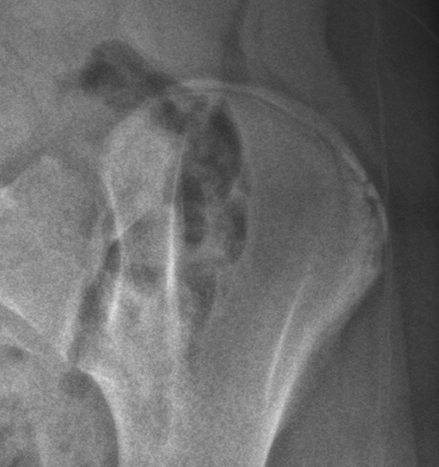
Background: Current brace weaning criteria for adolescents with idiopathic scoliosis (AIS) are not well defined. Risser Stage 4, ≥ 2 years since the onset of menarche, and no further increase in body height over 6 months are considered justifications for stopping bracing. However, despite adherence to such standards, curve progression still occurs in some patients, and so better criteria for brace discontinuation are needed.
Questions/purposes: (1) Is no change in height measurements over 6 months and Risser Stage 4 sufficient for initiating brace weaning? (2) What is the association between larger curves (45°) at brace weaning and the progression risk? (3) Are a more advanced Risser stage, Sanders stage, or distal radius and ulna classification associated with a decreased risk of curve progression? (4) When should we wean patients with AIS off bracing to reduce the time for brace wear while limiting the risk of postweaning curve progression?
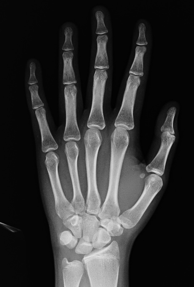
Methods: All AIS patients who were weaned off their braces from June 2014 to March 2016 were prospectively recruited and followed up for at least 2 years after weaning. A total of 144 patients were recruited with mean followup of 36 ± 21 months. No patients were lost to followup. Patients were referred for brace weaning based on the following criteria: they were Risser Stage 4, did not grow in height in the past 6 months of followup, and were at least 2 years postmenarche. Skeletal maturity was assessed with Risser staging, Sanders staging, and the distal radius and ulna classification. Curve progression was determined as any > 5° increase in the Cobb angle between two measurements from any subsequent six monthly followup visits. All radiographic measurements were performed by spine surgeons independently as part of their routine consultations and without knowledge of this study. Statistical analyses included an intergroup comparison of patients with and without curve progression, binomial stepwise logistic regression analysis, odds ratios (ORs) with their 95% confidence intervals (CIs), and a risk-ratio calculation. A reasonable protective maturity stage would generate an OR < 1.
Results: Among patients braced until they had no change in height for 6 months, were 2 years postmenarche for girls, and Risser Stage 4, 29% experienced curve progression after brace weaning. Large curves (≥ 45°) were associated with greater curve progression (OR, 5.0; 95% CI, 1.7-14.8; p = 0.002) as an independent risk factor. Patients weaned at Sanders Stage 7 (OR, 4.7; 95% CI, 2.1-10.7; p < 0.001), radius Grade 9 (OR, 3.9; 95% CI, 1.75-8.51; p = 0.001), and ulna Grade 7 (OR, 3.1; 95% CI, 1.27-7.38; p = 0.013) were more likely to experience curve progression. The earliest maturity indices with a reasonable protective association were Sanders Stage 8 (OR, 0.21; 95% CI, 0.09-0.48; p < 0.001), and radius Grade 10 (OR, 0.42; 95% CI, 0.19-0.97; p = 0.042) with ulna Grade 9 (no patients with curve progression).
Conclusion: Brace weaning indications using Risser staging are inadequate. Curve progression is expected in patients with large curves, irrespective of maturity status. Bone age measurement by either Sanders staging or the distal radius and ulna classification provides clearer guidelines for brace weaning, resulting in the least postweaning curve progression. Weaning in patients with Sanders Stage 8 and radius Grade 10/ulna Grade 9 provides the earliest and most protective timepoints for initiating brace weaning.
Level of evidence: Level II, prognostic study.
Conflict of interest statement
Each author certifies that neither he or she, nor any member of his or her immediate family, have funding or commercial associations (consultancies, stock ownership, equity interest, patent/licensing arrangements, etc) that might pose a conflict of interest in connection with the submitted article.
All ICMJE Conflict of Interest Forms for authors and
Figures


Comment in
-
CORR Insights®: When Should We Wean Bracing for Adolescent Idiopathic Scoliosis?Clin Orthop Relat Res. 2019 Sep;477(9):2158-2160. doi: 10.1097/CORR.0000000000000841. Clin Orthop Relat Res. 2019. PMID: 31283729 Free PMC article. No abstract available.
References
-
- Bitan FD, Veliskakis KP, Campbell BC. Differences in the Risser grading systems in the United States and France. Clin Orthop Relat Res . 2005:190-195. - PubMed
-
- Chang SH, Tzeng SJ, Cheng JY, Chie WC. Height and weight change across menarche of schoolgirls with early menarche. Arch Pediatr Adolesc Med . 2000;154:880-884. - PubMed
MeSH terms
LinkOut - more resources
Full Text Sources
Medical
Miscellaneous
