. 2019 Aug 15:333:576969.
doi: 10.1016/j.jneuroim.2019.576969. Epub 2019 May 20.

Imiquimod and interferon-alpha augment monocyte-mediated astrocyte secretion of MCP-1, IL-6 and IP-10 in a human co-culture system

Affiliations

Imiquimod and interferon-alpha augment monocyte-mediated astrocyte secretion of MCP-1, IL-6 and IP-10 in a human co-culture system

Michael D Rizzo et al. J Neuroimmunol. .

Abstract

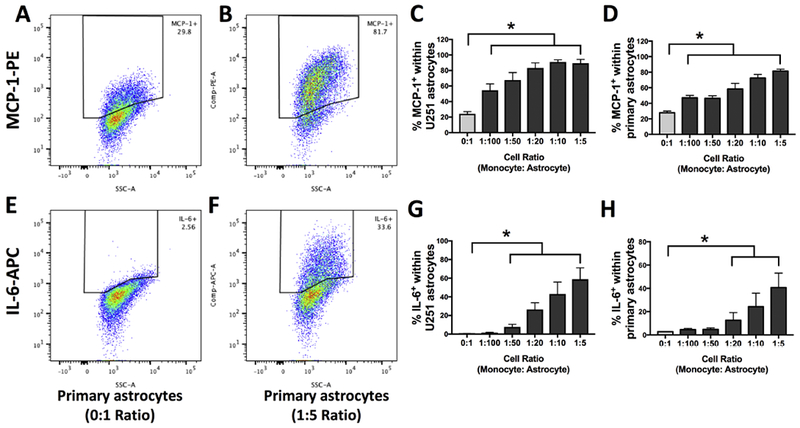
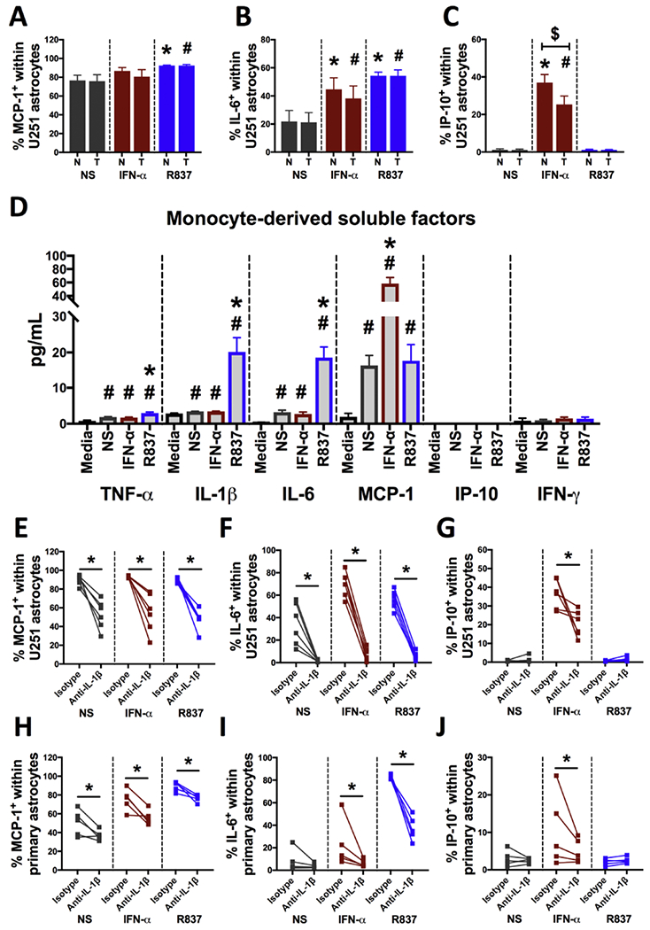
Toll-like receptor 7 (TLR7)-activation has been implicated as a significant mechanism of neuroinflammation triggered by ssRNA viruses. Infiltration of monocytes into the brain and astrocyte activation occurs during in vivo TLR7-mediated neuroinflammation. The objective here was to determine whether the TLR7 agonist, imiquimod, and interferon-alpha (IFN-α), promote monocyte-mediated astrocyte secretion of pro-inflammatory factors. Using a human primary co-culture system, we demonstrate that monocytes, together with imiquimod and IFN-α, promote astrocyte secretion of MCP-1, IL-6 and IP-10. Furthermore, TLR7-induced monocyte-derived IL-1β is critical for promoting the astrocyte response. Overall, this study provides a potential mechanism for TLR7-mediated neuroinflammation.

Keywords: Astrocyte; Co-culture; IL-1beta; Imiquimod; Interferon-alpha; Monocyte.


Conflict of interest statement

Competing interests: None of the authors present on this paper report any competing interests.

Figures

Figure 1:
Figure 1:. Human primary monocytes induce human U251 and primary astrocytes to produce MCP-1 and IL-6.
Human primary monocytes (N=5 for C-D and N=5 for G-H) were purified from healthy donors and co-cultured with U251 or primary astrocytes (donor 1). Astrocytes were cultured at 2×l05 cells/mL and monocytes were added at various concentrations to establish monocyte:astrocyte ratios. Cells were co-cultured for 24 h, with a Golgi block added 4 h before cell harvest. Intracellular staining and flow cytometry were performed to determine astrocyte production of MCP-1 and IL-6. FSC-A and FSC-H were used to separate the astrocyte population from the monocytes. Panels A-B and E-F are flow cytometry plots of astrocyte production of MCP-1 and IL-6 comparing astrocytes alone (0:1 ratio) to a monocyte to astrocyte ratio of 1:5. For statistical analysis, dataset was first log transformed. Next, a one-way (C and G) or repeated measures (RM) (D and H) analysis of variance (ANOVA) with a Dunnett’s multiple comparisons post-test was performed. * denotes a statistical difference from 0:1 ratio group (p<0.05). Graphs are mean +/− SEM.
Figure 2:
Figure 2:. Imiquimod/R837 treatment alone and with IFN-α, enhances monocyte-mediated astrocyte production of MCP-1, IL-6 and IP-10.
(A-F) Human primary monocytes were co-cultured with U251 or primary astrocytes (donor 1) at a ratio of 1:20 (monocyte:astrocyte) and stimulated with IFN-α (50, 100 and 200U/mL) or R837 (0.1, 1 and 10μg/mL). Cells were co-cultured for 24 h, with a Golgi block added 4 h before cell harvest. An aliquot of supernatant was collected prior to the addition of the Golgi block. Intracellular staining and flow cytometry were performed to determine astrocyte production of MCP-1, IL-6 and IP-10. The right panels in A-F are supernatant levels of cytokines in co-culture measured via LEGENDplex™ and/or ELISAmax™. The N values for monocytes isolated from human donors was N=9 and N=3 for left and right panels, respectively, in A, C, E, and N=5 for right and left panels in B, D, F. (G-L) IFN-α (100U/mL) and R837 (1μg/mL) were added alone and in combination to the co-culture and astrocyte production of MCP-1, IL-6 and IP-10 was determined (N=6 for G-I and N=5 for J-L). For A-F, dataset was log transformed and a RM ANOVA with a Dunnett’s multiple comparisons post-test was performed. For G-L, dataset was log transformed and a RM ANOVA with a Tukey’s post-test was performed. For A-F, * denotes a statistical difference from NS of the respective ratio group (p<0.05). Graphs are mean +/− SEM.
Figure 3:
Figure 3:. IL-1β is a major factor governing the monocyte-mediated astrocyte response to R837.
(A-C) Human monocytes (N=9) were co-cultured with U251 astrocytes at a ratio of 1:20 (monocyte:astrocyte) in either normal wells (N) or 0.4μm transwells (T). Co-culture was stimulated with IFN-α (100U/mL) or R837 (10μg/mL). Cells were co-cultured for 24 h, with a Golgi block added 4 h before cell harvest. (D) Human monocytes (N=9) were cultured alone at l×l04 cells/mL. Monocytes were either left untreated (NS), treated with IFN-α (100U/mL) or R837 (10μg/mL). Monocytes were cultured for 20 h and supernatants were collected. LEGENDplex™ was used to measure supernatant levels of MCP-1, TNF-α, IL-1β, IFN-γ, IL-6 and IP-10. (E-J) Human primary monocytes (N=6 for E-G and N=5 for H-J) were co-cultured with astrocytes (U251 or primary donor 1) at a ratio of 1:20 and stimulated with IFN-α (100U/mL) or R837 (10μg/mL). Anti-IL-1β was added to the co-culture at a concentration of 1μg/mL. An isotype (1 μg/mL) served as the control. Cells were co-cultured for 24 h, with a Golgi block added 4 h before cell harvest. For A-C, a RM two-way ANOVA with a Sidak’s multiple comparisons post-test was performed. * denotes a statistical difference from the NS group in normal (N) wells (p<0.05) and # denotes a statistical difference from the NS group in transwells (T). $ denotes a statistical difference between normal wells and transwells. For D, a log transformation was performed if there was unequal variance (F test). Then an unpaired t-test was performed to determine significant differences between media and each treatment. A RM ANOVA with a Dunnett’s multiple comparisons post-test was performed to determine differences between NS and either IFNα or R837-treated monocytes. # denotes a statistical difference between media control. * denotes a statistical difference from the NS (p<0.05). For E-J, a paired t-test was performed to determine significant differences between isotype control and anti-IL-1β group. A log transformation was performed for H-J prior to the paired t-test. * denotes a statistical difference from the isotype control (p<0.05). Graphs are mean +/− SEM.
Figure 4:
Figure 4:. Replacement of monocytes with recombinant IL-1β in the control, R837 and IFN-α cultures, promotes a similar profile of MCP-1, IL-6 and IP-10 production by astrocytes.
(A-F) Astrocytes (U251 and primary donor 1) were treated with varying concentrations of recombinant IL-1β (0, 0.5, 1, 5, 10, 20, 50 and 100pg/mL). (G-L) Astrocytes were left untreated (NS), treated with IFN-α (100U/mL) or R837 (10μg/mL) alone or in combination with IL-1β (20pg/mL). Cells were incubated for 20 h and a Golgi block was added for 4 h. Intracellular staining and flow cytometry were performed to determine astrocyte production of MCP-1, IL-6 and IP-10. A RM ANOVA with a Dunnett’s multiple comparisons post-test was performed for A-L. * denotes a statistical difference from 0pg/mL for A-F and the respective NS treatment group for G-L. Graphs are mean +/− SEM.
Figure 5:
Figure 5:. The intermediate monocyte population positively associates with U251 astrocyte production of IL-6.
Human primary monocytes (N=16) were co-cultured with U251 astrocytes at a ratio of 1:20 and stimulated with IFN-α (100U/mL) or R837 (10μg/mL). Cells were co-cultured for 24 h, with a Golgi blocker added 4 h before cell harvest. Intracellular staining and flow cytometry were performed to determine astrocyte production of MCP-1, IL-6 and IP-10. Extracellular staining of CD14 and CD16 was performed on an aliquot of monocytes prior to co-culture. The percent of each monocyte population was determined. A correlation analysis (Prism 7, GraphPad) was performed between monocyte subsets and U251 astrocyte production of MCP-1, IL-6 or IP-10.

References

    1. Abbott NJ, Ronnback L & Hansson E 2006. Astrocyte-endothelial interactions at the blood-brain barrier. Nat Rev Neurosci, 7, 41–53. - PubMed
    1. Aloisi F, Care A, Borsellino G, Gallo P, Rosa S, Bassani A, Cabibbo A, Testa U, Levi G & Peschle C 1992. Production of hemolymphopoietic cytokines (IL-6, IL-8, colony-stimulating factors) by normal human astrocytes in response to IL-1 beta and tumor necrosis factor-alpha. J Immunol, 149, 2358–66. - PubMed
    1. Anderson AM, Harezlak J, Bharti A, Mi D, Taylor MJ, Daar ES, Schifitto G, Zhong J, Alger JR, Brown MS, Singer EJ, Campbell TB, Mcmahon DD, Buchthal S, Cohen R, Yiannoutsos C, Letendre SL, Navia BA & Consortium HIVN 2015. Plasma and Cerebrospinal Fluid Biomarkers Predict Cerebral Injury in HIV-Infected Individuals on Stable Combination Antiretroviral Therapy. J Acquir Immune Defic Syndr, 69, 29–35. - PMC - PubMed
    1. Anderson AM, Lennox JL, Mulligan MM, Loring DW, Zetterberg H, Blennow K, Kessing C, Koneru R, Easley K & Tyor WR 2016. Cerebrospinal fluid interferon alpha levels correlate with neurocognitive impairment in ambulatory HIV-Infected individuals. J Neurovirol. - PMC - PubMed
    1. Andjelkovic AV, Kerkovich D & Pachter JS 2000. Monocyte:astrocyte interactions regulate MCP-1 expression in both cell types. JLeukoc Biol, 68, 545–52. - PubMed

Publication types

MeSH terms