. 2019 May 20;24(10):1940.
doi: 10.3390/molecules24101940.

The Characteristics of PD-L1 Inhibitors, from Peptides to Small Molecules

Affiliations

The Characteristics of PD-L1 Inhibitors, from Peptides to Small Molecules

Yanwen Zhong et al. Molecules. .

Abstract

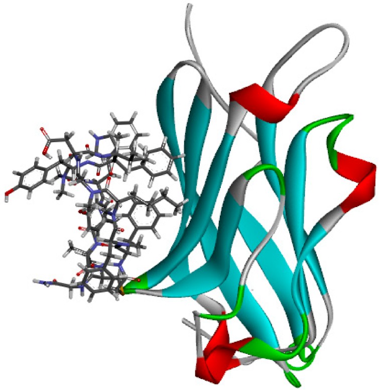
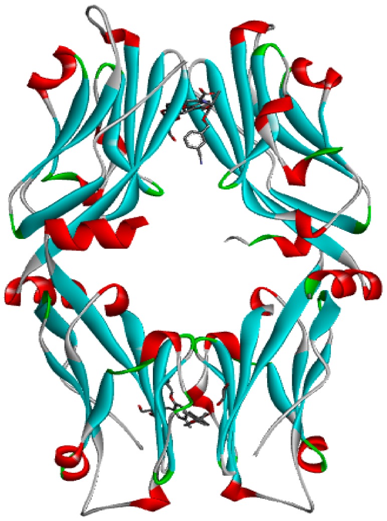
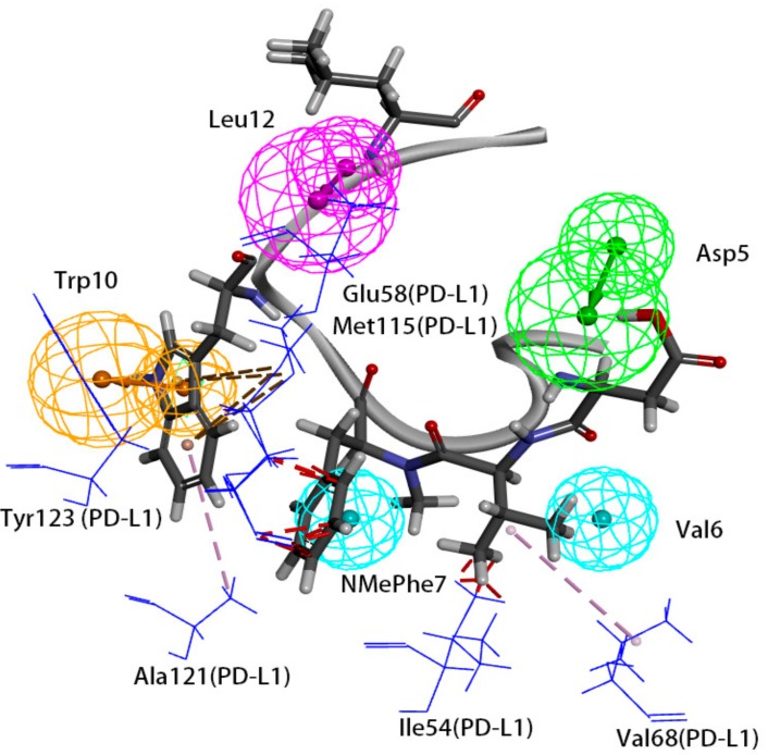
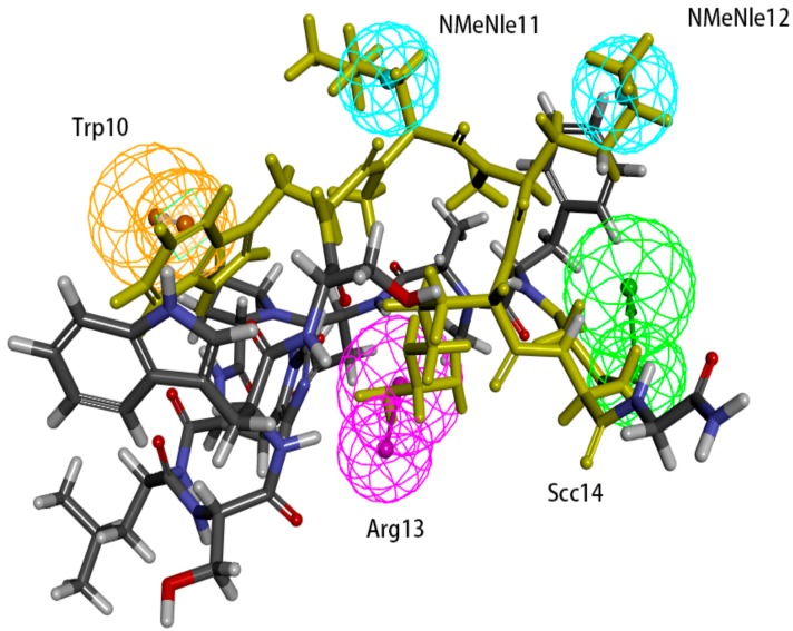
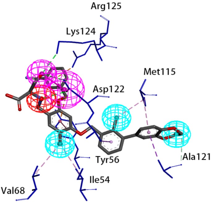
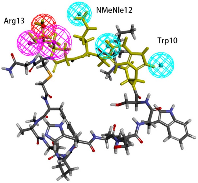
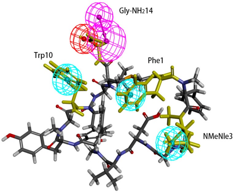
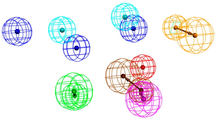
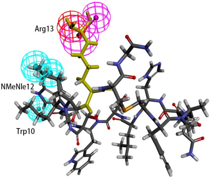
The programmed cell death ligand protein 1 (PD-L1) is a member of the B7 protein family and consists of 290 amino acid residues. The blockade of the PD-1/PD-L1 immune checkpoint pathway is effective in tumor treatment. Results: Two pharmacophore models were generated based on peptides and small molecules. Hypo 1A consists of one hydrogen bond donor, one hydrogen bond acceptor, two hydrophobic points and one aromatic ring point. Hypo 1B consists of one hydrogen bond donor, three hydrophobic points and one positive ionizable point. Conclusions: The pharmacophore model consisting of a hydrogen bond donor, hydrophobic points and a positive ionizable point may be helpful for designing small-molecule inhibitors targeting PD-L1.

Keywords: peptide; pharmacophore; programmed cell death ligand protein 1; small molecule.


Conflict of interest statement

The authors declare no conflict of interest.
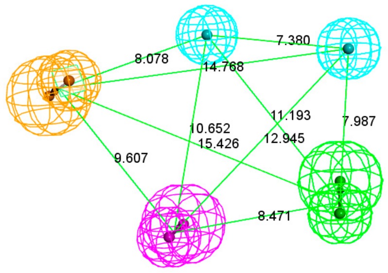
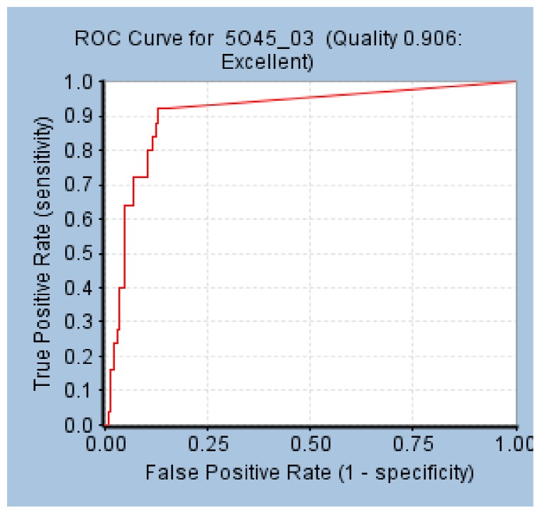
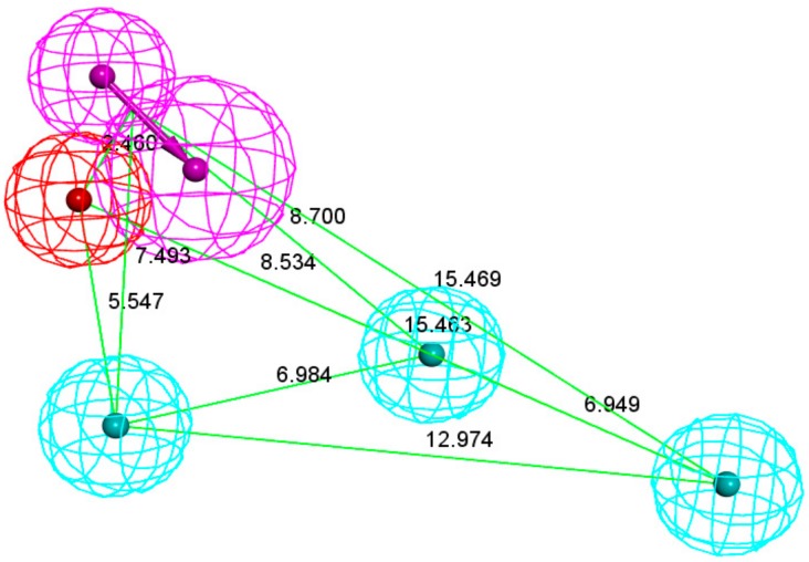
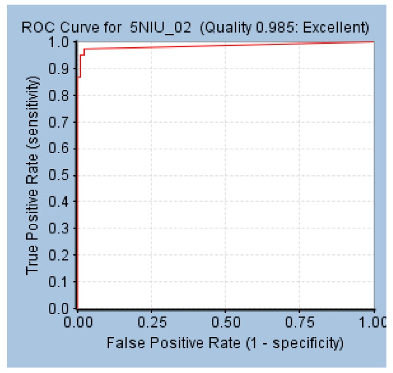
Figures

Figure 1
Figure 1
Pharmacophore model Hypo 1A. As the figure shows, the aromatic ring point is orange, the hydrophobic point is blue, the hydrogen bond acceptor is green, and the hydrogen bond donor is purple. The distance between the pharmacophore features is reported in angstroms.
Figure 2
Figure 2
Receiver operating characteristics (ROC) curve of Hypo 1A.
Figure 3
Figure 3
Pharmacophore model 1B. As the figure shows, the positive ionizable point is red, the hydrophobic point is blue, and the hydrogen bond donor is purple.
Figure 4
Figure 4
ROC curve of Hypo 1B.
Figure 5
Figure 5
The crystal structure of peptide-71 and human programmed cell death ligand 1 (hPD-L1) (Protein Data Bank (PDB) ID: 5O45).
Figure 6
Figure 6
The crystal structure of Bristol–Meyers–Squibb (BMS)-1001 and hPD-L1 (PDB ID: 5NIU).
Figure 7
Figure 7
Superposition between Hypo 1A and peptide-71. As the figure shows, the hydrophobic point is blue, the hydrogen bond donor is purple, the hydrogen bond acceptor is green, and the aromatic ring point is orange. The interacted residues of PD-L1 are blue.
Figure 8
Figure 8
Superposition between Hypo 1A and peptide-57. As the figure shows, the hydrophobic point is blue, the hydrogen bond donor is purple, the hydrogen bond acceptor is green, and the aromatic ring point is orange. The labelled residues are yellow.
Figure 9
Figure 9
Superposition between BMS-1001 and the pharmacophore model. As the figure shows, the positive ionizable point is red, the hydrophobic point is blue, and the hydrogen bond donor is purple. The interacted residues of PD-L1 are blue.
Figure 10
Figure 10
Superposition between Hypo 1B and peptide-57. As the figure shows, the positive ionizable point is red, the hydrophobic point is blue, and the hydrogen bond donor is purple. The labelled residues are yellow.
Figure 11
Figure 11
Superposition between Hypo 1B and peptide-71. As the figure shows, the positive ionizable point is red, the hydrophobic point is blue, and the hydrogen bond donor is purple. The labelled residues are yellow.
Figure 12
Figure 12
Superimposition of Hypo 1A and Hypo 1B. As the figure shows, in Hypo 1A, the hydrophobic point is light blue, the hydrogen bond acceptor is green, the hydrogen bond donor is purple, and the aromatic ring point is orange. In Hypo 1B, the hydrophobic point is Lyons blue, the positive ionizable point is red, and the hydrogen bond donor is brown.
Figure 13
Figure 13
Superposition between Hypo 1B and peptide-57. As the figure shows, the positive ionizable point is red, the hydrophobic point is blue, and the hydrogen bond donor is purple. The yellow amino acid is Arg13.

Similar articles

Cited by

References

    1. Massari F., Santoni M., Ciccarese C., Daniele S. The Immunocheckpoints in Modern Oncology: The next 15 Years. Expert Opin. Biol. Ther. 2015;15:917–921. doi: 10.1517/14712598.2015.1035251. - DOI - PubMed
    1. Bellmunt J., Powles T., Vogelzang N.J. A Review on the Evolution of Pd-1/Pd-L1 Immunotherapy for Bladder Cancer: The Future is Now. Cancer Treat. Rev. 2017;54:58–67. doi: 10.1016/j.ctrv.2017.01.007. - DOI - PubMed
    1. Boussiotis V.A. Molecular and Biochemical Aspects of The Pd-1 Checkpoint Pathway. New Engl. J. Med. 2016;375:1767–1778. doi: 10.1056/NEJMra1514296. - DOI - PMC - PubMed
    1. Collin M. Immune Checkpoint Inhibitors: A Patent Review (2010–2015) Expert Opin. Ther. Patents. 2016;26:555–564. doi: 10.1080/13543776.2016.1176150. - DOI - PubMed
    1. Wajant H. Death Receptors. Essays Biochem. 2003;39:53–71. doi: 10.1042/bse0390053. - DOI - PubMed

MeSH terms