. 2019 May 27;20(10):2597.
doi: 10.3390/ijms20102597.

Engineering of Nanobodies Recognizing the Human Chemokine Receptor CCR7

Affiliations

Engineering of Nanobodies Recognizing the Human Chemokine Receptor CCR7

Barbara D Jakobs et al. Int J Mol Sci. .

Abstract

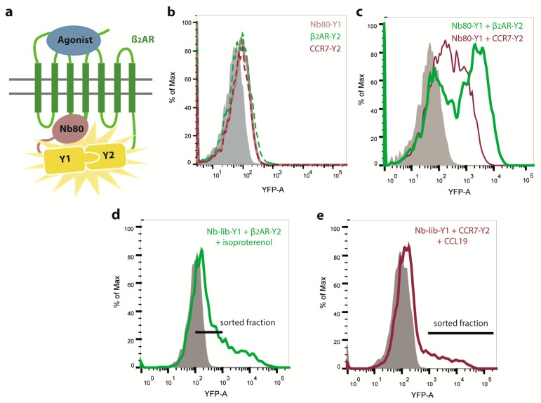
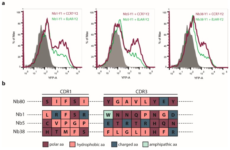
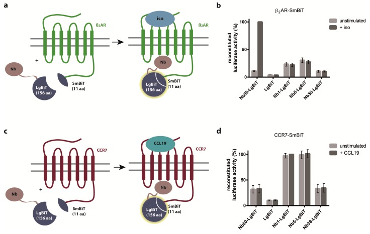
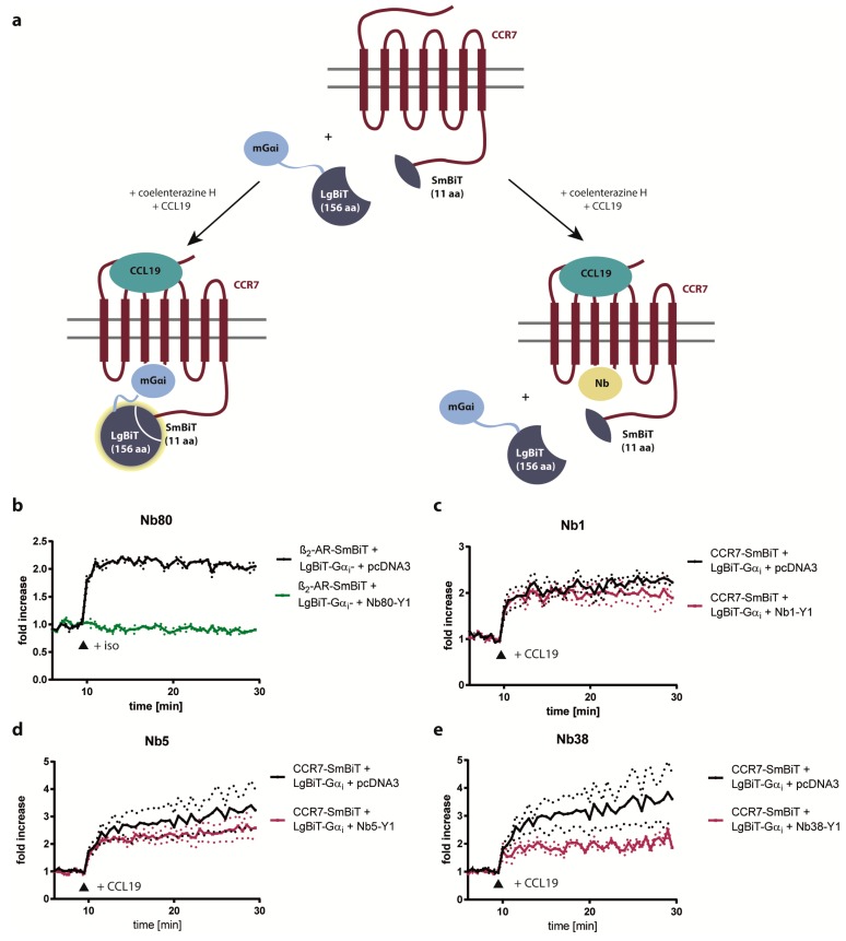
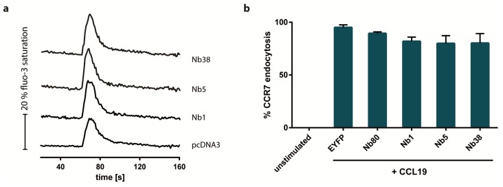
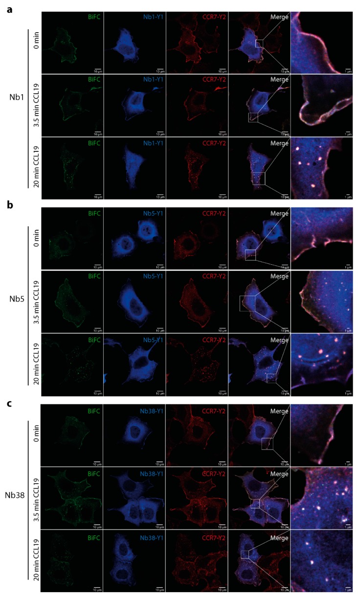
The chemokine receptor CCR7 plays a pivotal role in health and disease. In particular, CCR7 controls homing of antigen-bearing dendritic cells and T cells to lymph nodes, where adaptive immune responses are initiated. However, CCR7 also guides T cells to inflamed synovium and thereby contributes to rheumatoid arthritis and promotes cancer cell migration and metastasis formation. Nanobodies have recently emerged as versatile tools to study G-protein-coupled receptor functions and are being tested in diagnostics and therapeutics. In this study, we designed a strategy to engineer novel nanobodies recognizing human CCR7. We generated a nanobody library based on a solved crystal structure of the nanobody Nb80 recognizing the β2-adrenergic receptor (β2AR) and by specifically randomizing two segments within complementarity determining region 1 (CDR1) and CDR3 of Nb80 known to interact with β2AR. We fused the nanobody library to one half of split-YFP in order to identify individual nanobody clones interacting with CCR7 fused to the other half of split-YFP using bimolecular fluorescence complementation. We present three novel nanobodies, termed Nb1, Nb5, and Nb38, that recognize human CCR7 without interfering with G-protein-coupling and downstream signaling. Moreover, we were able to follow CCR7 trafficking upon CCL19 triggering using Nb1, Nb5, and Nb38.

Keywords: CCL19; CCR7; bimolecular fluorescence complementation; chemokine receptor; chemokines; nanobodies; split-luciferase complementation; β2-adrenergic receptor.


Conflict of interest statement

The authors declare no conflict of interest. The funders had no role in the design of the study; in the collection, analyses, or interpretation of data; in the writing of the manuscript, or in the decision to publish the results.

Figures

Figure 1
Figure 1
Engineering of nanobodies (Nbs) recognizing CCR7 by bimolecular fluorescence complementation (BiFC). Structure and sequence of the conformation-specific Nb80, which recognizes an isoproterenol-activated conformation of the β2-adrenergic receptor (β2AR), was used to specifically randomize complementarity determining region (CDR)1 and CDR3 to generate a new Nb library. The newly generated Nb library was fused to the N-terminal part of split-YFP (YFP1, Y1) in order to identify Nbs that recognize CCR7 fused to split-YFP2 (Y2) by BiFC. (a) Schematic illustration of BiFC between Nb80-YFP1 and β2AR-YFP2. Nb80 recognizes and binds to agonist activated β2AR. Thereby, the two split-YFP fragments will reconstitute to form native YFP. (b) Flow cytometric analysis of HEK293 cells transiently expressing either Nb80-YFP1 (dotted brown line), β2AR-YFP2 (dotted green line), or CCR7-YFP2 (dotted red line) alone. Untransfected, control cells are shown in grey. (c) Flow cytometric analysis showing BiFC in HEK293 cells transiently co-expressing Nb80-YFP1 and β2AR-YFP2 (green line) as proof of concept or Nb80-YFP1 and CCR7-YFP2 (red line) as control. Before measuring YFP fluorescence, cells were stimulated with isoproterenol (10 µM) or CCL19 (0.5 µg/mL), respectively. (d) Negative screening of Nb library to remove β2AR-recognizing Nbs. HEK293 cells were transiently transfected with the newly generated Nb library fused to split-YFP1 (Nb-lib-Y1) and β2AR fused to split-YFP2. After isoproterenol stimulation (10 µM), BiFC-negative cells were FACS sorted to enrich the Nb library for Nbs that do not interact with β2AR anymore. Sorted cell fraction is indicated by the black line. Afterwards, plasmids coding for the Nb library were isolated. (e) BiFC of remaining Nb library-YFP1 and CCR7-YFP2. The Nb library-YFP1 was transiently expressed in HEK293 cells stably expressing CCR7-YFP2 and cells were stimulated with CCL19 (0.5 µg/mL). BiFC-positive cells, indicated by the black line, were FACS sorted.
Figure 2
Figure 2
Flow cytometric analysis (a) and sequence analysis (b) of individual Nb clones previously selected by BiFC screening. (a) Plasmids coding for Nb-YFP1 (Y1) were isolated and individual clones were co-transfected again with CCR7-YFP2 (Y2) to screen for single Nb clones that recognize CCR7. Here, the three most promising Nb clones are represented: Nb1, Nb5, and Nb38. BiFC between individual Nb clones and CCR7 is indicated in red. Additionally, BiFC of individual Nb-YFP1 clones and β2AR-YFP2 was analyzed and is depicted in green. (b) Nb1, Nb5, and Nb38 were sequenced. Protein sequences of CDR1 and CDR3 of each Nb are illustrated in comparison to Nb80. Different colors were used to highlight characteristic properties of respective amino acids (aa).
Figure 3
Figure 3
Nb80 preferentially interacts with active β2AR, whereas Nb1, Nb5, and Nb38 preferentially recognize CCR7 independent of its activation state as assessed by split-luciferase complementation. (a,c) Schematic representation of the split-luciferase complementation assay. Nb-GPCR interactions are determined by reconstitution of Small BiT (SmBiT) and Large BiT (LgBiT) to functional NanoLuc (NLuc) luciferase before and after agonist stimulation and subsequent measurements of luminescence signals generated by the reconstituted luciferase. (b,d) HEK293 cells transiently co-expressing β2AR (b) or CCR7 (d) fused to SmBiT of NLuc and individual Nb clones fused to LgBiT of NLuc were incubated with coelenterazine H (5µM), the luciferase’s substrate, and after 10 min, stimulated with isoproterenol (iso) (10 µM) (b) or CCL19 (1.5 µg/mL) (d). As control, we transiently co-expressed LgBiT without Nb together with either GPCR-SmBiT. Reconstituted luciferase activity between Nb80 and β2AR (b) and Nb1 and CCR7 (d), respectively, was set to 100%. Results represent each the mean values of three independent experiments including the standard error of the mean (SEM).
Figure 4
Figure 4
CCL19-triggered G-protein coupling to CCR7 is barely impaired by Nb1, Nb5, or Nb38. (a) Schematic illustration of the G-protein competition assay based on split-luciferase complementation. (be) HEK293 cells transiently expressing β2AR-SmBiT (b) or CCR7-SmBiT (ce) together with LgBiT-mGαi and Nb-YFP1 (Nb-Y1) constructs were incubated with coelenterazine H (5 µM) and subsequently stimulated (indicated by the arrow head) either with 10 µM isoproterenol (b) or 1.5 µg/mL CCL19 (ce), respectively. As control, HEK293 cells were transiently co-transfected with GPCR-SmBiT, LgBiT-mGαi and pcDNA3 (indicated in black). In this case, increase in luminescence indicated functional activity of reconstituted split NLuc luciferase upon recruitment and interaction of mGαi with the GPCR. Replacing empty vector (pcDNA3) with Nb80-YFP1 (b) caused complete blockage in luciferase activity (indicated in green). Expressing Nb1-YFP1 (c), Nb5-YFP1 (d) or Nb38-YFP1 (e) barely interfered with mGαi-coupling to CCR7 (indicated in red). Results represent the fold increase in luminescence over the baseline, which is set to 1, upon agonist-stimulation. Mean values and SEM of three independent experiments are shown.
Figure 5
Figure 5
CCL19-mediated calcium mobilization and CCR7 endocytosis are not affected by Nb1, Nb5, or Nb38. (a) Influence of intracellular expression of the CCR7-recognizing Nbs, Nb1, Nb5, and Nb38, on CCL19-mediated changes in intracellular calcium concentrations was measured in H1299 cells stably expressing CCR7-HA. Cells were stimulated with 0.5 µg/mL of CCL19. A representative experiment out of three is shown. (b) Endocytosis of CCR7 triggered by 1 µg/mL CCL19 was analyzed in HEK293-CCR7-HA cells transiently expressing CCR7-recognizing Nbs. Co-expressing EYFP or Nb80 instead of CCR7-recognizing Nbs served as controls. Mean values and SEM out of three independent experiments are presented.
Figure 6
Figure 6
Monitoring CCR7 trafficking triggered by CCL19 using Nb1, Nb5, and Nb38. Interaction between CCR7-YFP2 (Y2) and individual Nbs fused to YFP1 was assessed by BiFC and confocal microscopy in HEK293 transfected cells that were unstimulated or stimulated for indicated time points with 0.5 µg/mL of CCL19. Nb1-YFP1 (Y1) (a), Nb5-YFP1 (Y1) (b), and Nb38-YFP1 (Y1) (c). In addition, Nb-Y1 and CCR7-Y2 were immunostained using anti-YFP1 and anti-YFP2 specific antibodies. Scale bar: 10 µm, zoomed image: 1 µm.

References

    1. Griffith J.W., Sokol C.L., Luster A.D. Chemokines and Chemokine Receptors: Positioning Cells for Host Defense and Immunity. Annu. Rev. Immunol. 2014;32:659–702. doi: 10.1146/annurev-immunol-032713-120145. - DOI - PubMed
    1. Legler D., Thelen M. Chemokines: Chemistry, Biochemistry and Biological Function. Chim. Int. J. Chem. 2016;70:856–859. doi: 10.2533/chimia.2016.856. - DOI - PubMed
    1. Laufer J.M., Legler D.F. Beyond migration—Chemokines in lymphocyte priming, differentiation, and modulating effector functions. J. Leukoc. Biol. 2018;104:301–312. doi: 10.1002/JLB.2MR1217-494R. - DOI - PubMed
    1. Marcuzzi E., Angioni R., Molon B., Cal B. Chemokines and Chemokine Receptors: Orchestrating Tumor Metastasization. Int. J. Mol. Sci. 2018;20:96. doi: 10.3390/ijms20010096. - DOI - PMC - PubMed
    1. Kufareva I., Salanga C.L., Handel T.M., Diego S., Jolla L. Chemokine and chemokine receptor structure and interactions: implications for therapeutic strategies. Immunol. Cell Biol. 2015;93:372–383. doi: 10.1038/icb.2015.15. - DOI - PMC - PubMed

MeSH terms