Review
. 2019 May 27;11(5):1186.
doi: 10.3390/nu11051186.

Nutritional Strategies to Prevent Lens Cataract: Current Status and Future Strategies

Affiliations
Review

Nutritional Strategies to Prevent Lens Cataract: Current Status and Future Strategies

Andrea J Braakhuis et al. Nutrients. .

Abstract

Oxidative stress and the subsequent oxidative damage to lens proteins is a known causative factor in the initiation and progression of cataract formation, the leading cause of blindness in the world today. Due to the role of oxidative damage in the etiology of cataract, antioxidants have been prompted as therapeutic options to delay and/or prevent disease progression. However, many exogenous antioxidant interventions have to date produced mixed results as anti-cataract therapies. The aim of this review is to critically evaluate the efficacy of a sample of dietary and topical antioxidant interventions in the light of our current understanding of lens structure and function. Situated in the eye behind the blood-eye barrier, the lens receives it nutrients and antioxidants from the aqueous and vitreous humors. Furthermore, being a relatively large avascular tissue the lens cannot rely of passive diffusion alone to deliver nutrients and antioxidants to the distinctly different metabolic regions of the lens. We instead propose that the lens utilizes a unique internal microcirculation system to actively deliver antioxidants to these different regions, and that selecting antioxidants that can utilize this system is the key to developing novel nutritional therapies to delay the onset and progression of lens cataract.

Keywords: antioxidant supplements; cataract; dietary antioxidants; lens.


Conflict of interest statement

The authors declare no conflict of interest.

Figures

Figure 1
Figure 1
Cortical cataracts. (A) Location of the cortical cataract subtype. Top panel: diagram showing the opacities that form in the lens cortex. Lower panel: Scheimpflug slit-lamp photographic image revealing a cortical cataract. (B) Molecular mechanisms involved in the pathogenesis of diabetic cortical cataract. An increase in glucose leads to a decrease in GSH and an increase in reactive oxygen species (ROS) as indicated by the red arrows. The induced osmotic and oxidative stress work synergistically to inhibit the ability of fibre cells to regulate their volume. This leads to cell swelling, depolarization and an influx of sodium and calcium ions. The accumulation of calcium ions results in the activation of calcium-dependent proteases, which target cytoskeletal and crystallin proteins. Furthermore, proteins are modified by the formation of advanced glycation end (AGEs) products, which are known to alter the structure and function of crystallins, resulting in an increase in insoluble proteins, the formation of high molecular weight aggregates, and cataract.
Figure 2
Figure 2
Nuclear cataracts. (A) Location of the nuclear cataract subtype. Left panel: diagram showing the opacities that form in the lens nucleus. Right panel: Scheimpflug slit-lamp photographic image revealing a nuclear cataract. (B) Molecular mechanisms involved in the pathogenesis of age-related nuclear cataract. GSH levels are maintained at high levels within the lens by a combination of pathways including regeneration of oxidised GSH (GSSG) back to GSH via the enzyme glutathione reductase (GR) as well as repair enzymes thioltransferase (Ttase), which dethiolate protein mixed disulfides, such as protein bound GSH (PSSG), and thioredoxin (TrX), that dethiolates protein-protein disulphides (PSSP). In age related nuclear cataract, depletion of GSH levels in the nucleus, but not the lens cortex, results in significant oxidation of nuclear proteins, an increase in protein mixed disulphides, formation of protein-protein disulfide bonds, protein aggregation, loss of protein solubility, increased yellowing of the lens nucleus, and eventual nuclear cataract formation.
Figure 3
Figure 3
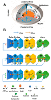
Lens structure and function (A) 3-D representation of the microcirculation model, showing ions and fluid fluxes that enter the lens at both poles via the extracellular space (blue arrows) before crossing fiber cell membranes and exiting the lens at the equator via an intracellular pathway (red arrows) mediated by gap junctions. (B) Equatorial cross-sections showing how the spatial differences in the distribution of ion channels and transporters between the epithelium (E), differentiating (DF) and mature (MF) fiber cells that generate the circulating flux of Na+ ions (top) that drives isotonic fluid fluxes (middle) which in turn deliver nutrients to and remove metabolic waste from the MF cells (bottom).

References

    1. Hobbs R.P., Bernstein P.S. Nutrient Supplementation for Age-related Macular Degeneration, Cataract, and Dry Eye. J. Ophthalmic Vis. Res. 2014;9:487–493. doi: 10.4103/2008-322X.150829. - DOI - PMC - PubMed
    1. Pascolini D., Mariotti S.P. Global estimates of visual impairment: 2010. Br. J. Ophthalmol. 2012;96:614–618. doi: 10.1136/bjophthalmol-2011-300539. - DOI - PubMed
    1. Weikel K.A., Garber C., Baburins A., Taylor A. Nutritional modulation of cataract. Nutr. Rev. 2014;72:30–47. doi: 10.1111/nure.12077. - DOI - PMC - PubMed
    1. Wild S., Roglic G., Green A., Sicree R., King H. Global prevalence of diabetes: estimates for the year 2000 and projections for 2030. Diabetes Care. 2004;27:1047–1053. doi: 10.2337/diacare.27.5.1047. - DOI - PubMed
    1. Klein B.E., Klein R., Wang Q., Moss S.E. Older-onset diabetes and lens opacities. The Beaver Dam Eye Study. Ophthalm. Epid. 1995;2:49–55. doi: 10.3109/09286589509071451. - DOI - PubMed