. 2019 May 28;7(1):140.
doi: 10.1186/s40425-019-0622-0.

Generation of monocyte-derived tumor-associated macrophages using tumor-conditioned media provides a novel method to study tumor-associated macrophages in vitro

Affiliations

Generation of monocyte-derived tumor-associated macrophages using tumor-conditioned media provides a novel method to study tumor-associated macrophages in vitro

Brooke Benner et al. J Immunother Cancer. .

Abstract

Background: Tumor-associated macrophages (TAM) are expanded and exhibit tumor-promoting properties within the tumor microenvironment. Current methods to study TAM have not been replicated across cancer types and often do not include exogenous growth factors from the tumor, a key factor in TAM differentiation in vivo.

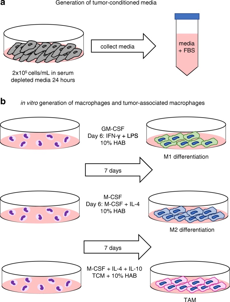
Methods: In this study, an in vitro method to generate monocyte- derived TAM using tumor- conditioned media (TCM) and a cytokine cocktail containing IL-4, IL-10, and M-CSF was utilized to study the phenotype, morphology, and function of TAM across multiple cancer types. TCM was generated from two breast cancer cell lines and an Epstein-Barr virus-positive lymphoma cell line. The properties of in vitro generated TAM were compared to in vitro generated M1 and M2- like macrophages and TAM isolated from patients with cancer.

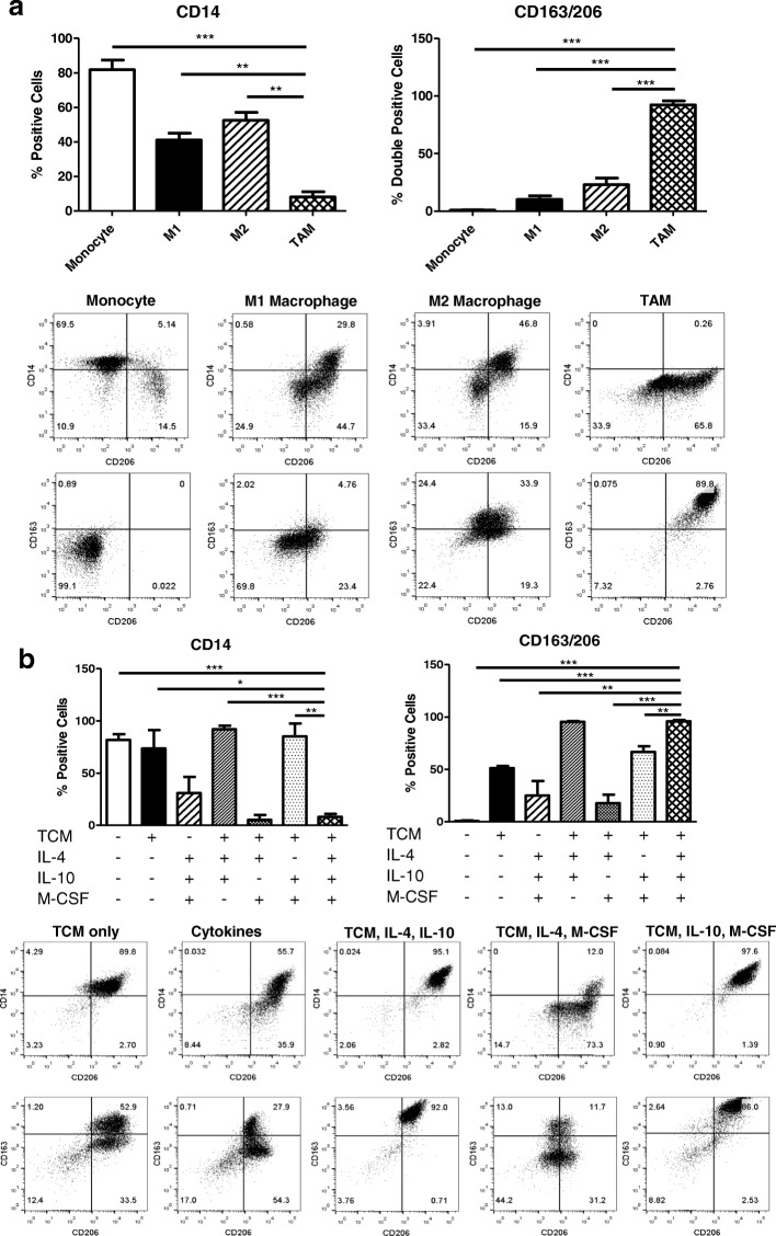
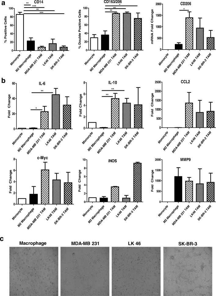
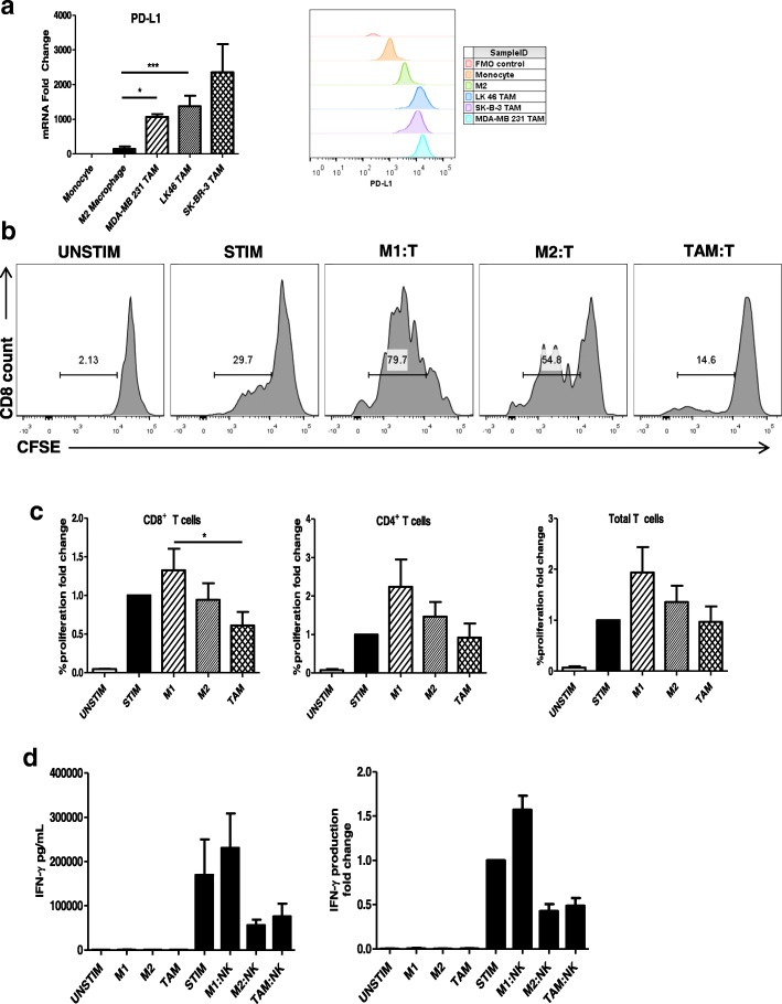
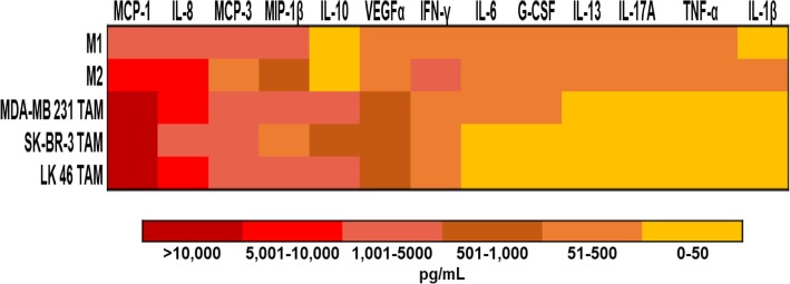
Results: TAM generated in this fashion displayed an increase in CD163/CD206 co-expression compared to M2- like macrophages (87 and 36%, respectively). TAM generated in vitro exhibited increased transcript levels of the functional markers IL-6, IL-10, CCL2, c-Myc, iNOS, and arginase compared to in vitro generated M2-like macrophages. Functionally, in vitro generated TAM inhibited the proliferation of T cells (47% decrease from M1-like macrophages) and the production of IFN-γ by natural killer cells was inhibited (44%) when co-cultured with TAM. Furthermore, in vitro generated TAM secreted soluble factors that promote the growth and survival of tumor cells.

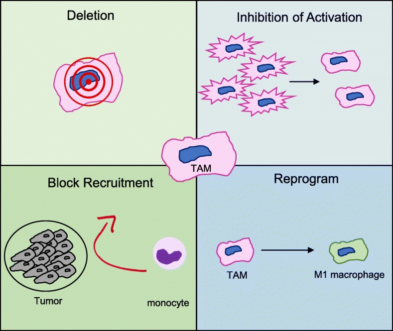
Conclusions: Limited access to patient TAM highlights the need for methods to generate TAM in vitro. Our data confirm that monocyte-derived TAM can be generated reliably using TCM plus the cytokine cocktail of IL-4, IL-10, and M-CSF. Given the ability of TAM to inhibit immune cell function, continued study of methods to deplete or deactivate TAM in the setting of cancer are warranted.

Keywords: Cancer; Immunotherapy; In vitro generation; Tumor-associated macrophages.


Conflict of interest statement

The authors declare that they have no competing interests.

Figures

Fig. 1
Fig. 1
Generation of tumor-conditioned media and in vitro macrophages. (a) Schematic of the generation of tumor-conditioned media (TCM) using 2 × 10⁵ cells/mL (MDA- MB 231, SK-BR-3 or LK 46) in 0.2% FBS media for 24 h. Media was collected and centrifuged to remove any tumor cells. 10% FBS was added. TCM was stored in 5 mL aliquots in − 80° freezer until use. (b) Schematic of the in vitro generation of macrophages. Monocytes harvested from healthy donor blood were plated at 1 × 10⁶ cells/mL in a total of 10 mL 10% HAB (M1/M2) or 5 mL 10% HAB plus 5 mL TCM (TAM). In M1-like monocyte differentiation, GM-CSF (1 μg/mL) was added to culture for 6 days. On day 6 the media was refreshed with GM-CSF (1 μg/mL) + IFN-γ (20 ng/mL) + LPS (50 ng/mL) for 24 h. In M2-like differentiation, M-CSF (1 μg/mL) was added to culture for 6 days. On day 6 the media was refreshed with M-CSF (1 μg/mL) and IL-4 (1 μg/mL). In TAM differentiation, M-CSF, IL-10, and IL-4 (all at 1 μg/mL) were added to culture in combination with TCM and 10% HAB (1:1). In all differentiation protocols media was refreshed every other day and cells were harvested on day 7
Fig. 2
Fig. 2
Characterization of in vitro generation methods of tumor-associated macrophages. (a) In vitro generated TAM were compared to classically and alternatively activated macrophages. Cells were stained on generation day 7 to assess surface expression of CD14, CD163, and CD206 analyzed by flow cytometry. Representative flow plots are shown. (b) The method used to generate TAM in vitro was validated using 6 conditions (TCM only, cytokines only, TCM + IL-4 + IL-10, TCM + IL-4 + M-CSF, TCM + IL-10 + M-CSF, TCM + IL- 4 + IL- 10 + M-CSF). Surface expression of CD14, CD163, and CD206 was measured on day 7 and analyzed by flow cytometry. Representative flow plots are shown
Fig. 3
Fig. 3
Phenotype and functional characterization of in vitro generated TAM. (a) M2-like macrophages and in vitro generated TAM were flow stained for surface expression of CD14 and CD163/CD206 co-expression and were analyzed by real-time PCR for transcript expression of CD206. (b) M2-like macrophages and in vitro generated TAM were analyzed by real-time PCR for transcript expression of IL-6 (macrophage-associated inflammatory pathway), IL-10 (immunosuppressive cytokine), CCL2 (macrophage stimulating chemokine), c-Myc (pro-tumor oncogene), iNOS (inducible nitric oxide synthase; tumor-protecting [54]), and MMP9 (matrix metallopeptidase 9; mediated extracellular matrix degradation and release of bioactive VEGFα [55]). (c) Brightfield images of in vitro generated macrophages and TAM generated using TCM from different tumor types at day 7
Fig. 4
Fig. 4
Human-derived TAM phenotype. (a) Melanoma and normal adjacent tissue were digested, stained for CX3CR1, CD163, and CD206 and analyzed by flow cytometry. Cell populations were first gated on the CD45+ and lineage (CD3, CD19, CD56) population. (b) SK-BR-3 in vitro generated TAMs were stained with antibodies for CX3CR1, CD163, and CD206 and analyzed by flow cytometry. Cell populations were first gated on the CD45+ and lineage (CD3, CD19, CD56) population
Fig. 5
Fig. 5
in vitro generated TAM inhibit T cell and NK cell function. (a) M2-like macrophages and in vitro generated TAM were analyzed by real-time PCR for transcript expression and flow cytometry for surface expression of PD-L1 CD3/CD28 bead activated T cells labelled with CFSE were co-cultured with M1-like and M2-like macrophages and MDA-MB 231 conditioned in vitro generated TAM at a 1:1 ratio. After 72 h cells were collected and (b) proliferation was evaluated by flow cytometry. Histograms show results from one representative experiment of CD8+ T cells. (c) Percent T cell proliferation expressed as a fold change and percentage. Bars represent the mean concentration ± SD of T cell proliferation analyzed by flow cytometry. Data are representative of three independent experiments with similar results; p < 0.05. (d) NK cells stimulated with IL-12, IL-18 and immobilized IgG were co-cultured with M1-like and M2- like macrophages and MDA-MB 231-conditioned in vitro generated TAM at a 1:1 ratio for 72 h. Bars represent the mean concentration ± SD of IFN-γ content from culture supernatants analyzed by ELISA. Data are representative of three independent experiments with similar results
Fig. 6
Fig. 6
in vitro generated TAM secrete growth factors that support the growth and survival of tumor cells. In vitro generated TAM secrete inflammatory cytokines. Culture supernatants were harvested from M1 and M2-like macrophages and in vitro generated TAM conditioned from MDA-MB 231, SK- BR-3, and LK 46 tumor-conditioned media. Data are presented as a heat map for expression of soluble factors in supernatants from each macrophage phenotype
Fig. 7
Fig. 7
Therapeutic targeting of tumor-associated macrophages in cancer. TAM are a promising target for the immunotherapy of cancer. Several approaches to reduce TAM or functionally modify TAM include blocking monocyte recruitment to the tumor, deletion through antigen-specific targeting, reprogramming of TAM to M1 macrophages and inhibiting the activation of TAM

References

    1. Fukuda K, Kobayashi A, Watabe K. The role of tumor-associated macrophage in tumor progression. Front Biosci (Schol Ed) 2012;4:787–798. - PubMed
    1. Liu Y, Cao X. The origin and function of tumor-associated macrophages. Cell Mol Immunol. 2015;12:1–4. doi: 10.1038/cmi.2014.83. - DOI - PMC - PubMed
    1. Gajewski TF, Schreiber H, Fu Y-X. Innate and adaptive immune cells in the tumor microenvironment. Nat Immunol. 2013;14:1014–1022. doi: 10.1038/ni.2703. - DOI - PMC - PubMed
    1. Stiff A, Trikha P, Wesolowski R, et al. Myeloid-derived suppressor cells express Bruton’s tyrosine kinase and can be depleted in tumor-bearing hosts by Ibrutinib treatment. Cancer Res. 2016;76:2125–2136. doi: 10.1158/0008-5472.CAN-15-1490. - DOI - PMC - PubMed
    1. Noy R, Pollard JW. Tumor-associated macrophages: from mechanisms to therapy. Immunity. 2014;41:49–61. doi: 10.1016/J.IMMUNI.2014.06.010. - DOI - PMC - PubMed

Publication types

Substances