. 2019 Sep;17(9):1920-1930.
doi: 10.1158/1541-7786.MCR-18-1291. Epub 2019 May 28.

Serine/Arginine-Rich Splicing Factor 3 Modulates the Alternative Splicing of Cytoplasmic Polyadenylation Element Binding Protein 2

Affiliations

Serine/Arginine-Rich Splicing Factor 3 Modulates the Alternative Splicing of Cytoplasmic Polyadenylation Element Binding Protein 2

James T DeLigio et al. Mol Cancer Res. 2019 Sep.

Abstract

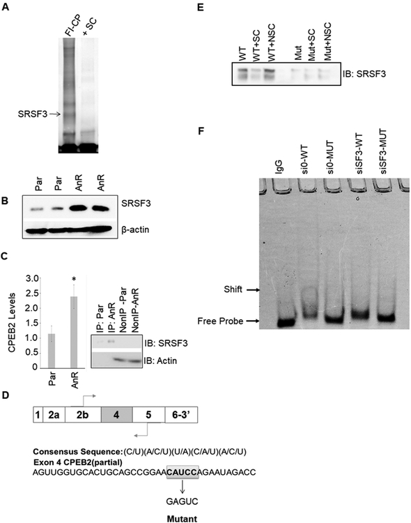
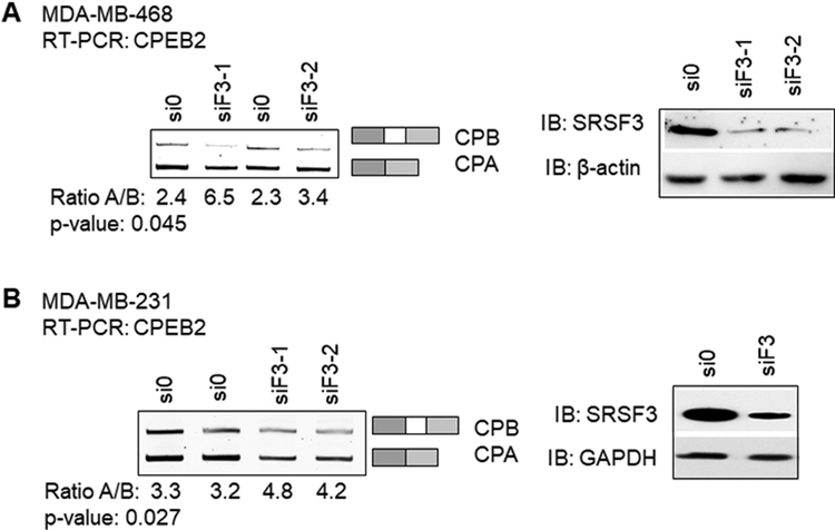
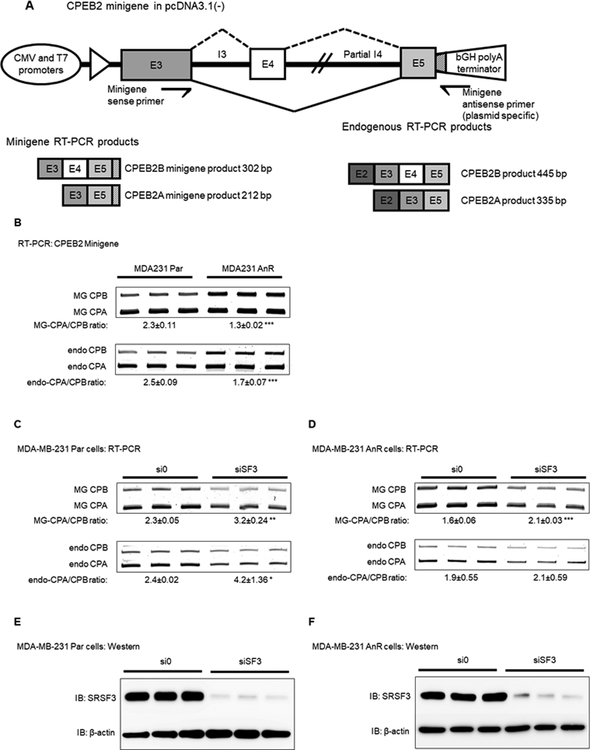
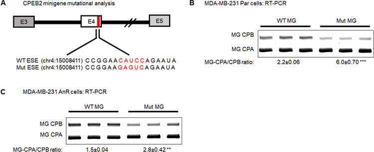
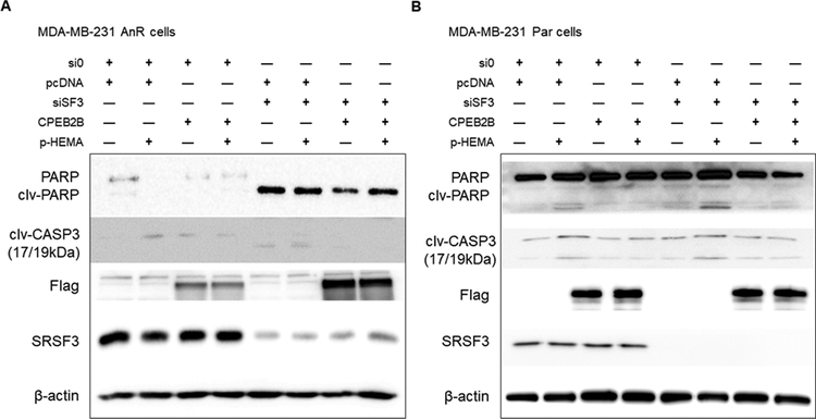
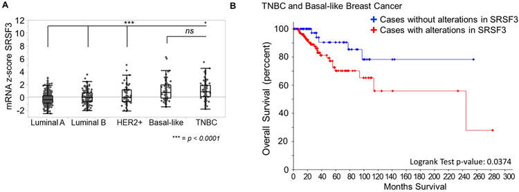
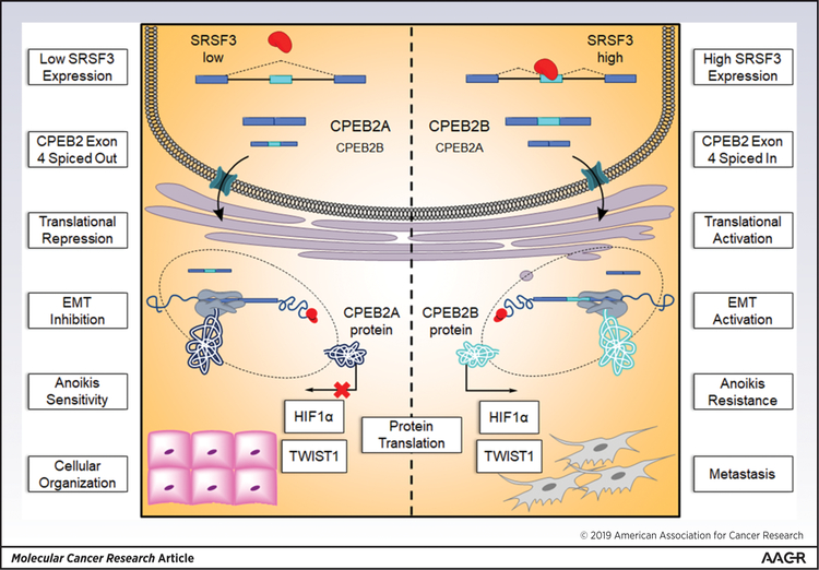
Triple negative breast cancer (TNBC) has an unusually low 5-year survival rate linked to higher metastatic rates. Our laboratory recently delineated a role for the alternative RNA splicing (AS) of cytoplasmic polyadenylation element binding protein 2 (CPEB2), via inclusion/exclusion of exon 4, in the metastasis of TNBC. In these studies, the mechanism governing the inclusion/exclusion of exon 4 was examined. Specifically, the RNA trans-factor, SRSF3, was found to be explicitly associated with CPEB2 exon 4. A SRSF3 consensus sequence was identified in exon 4, and mutation of this sequence abolished the association of SRSF3. The expression of SRSF3 was upregulated in TNBC cells upon the acquisition of anoikis resistance correlating with a reduction in the CPEB2A/B ratio. Importantly, downregulation of SRSF3 in these cells by siRNA induced the exclusion of exon 4 in cells increasing the ratio of CPEB2A (exon 4 excluded) to CPEB2B (exon 4 included). Downregulation of SRSF3 also reversed the CPEB2A/B ratio of a wild-type CPEB2 exon 4 minigene and endogenous CPEB2 pre-mRNA, but not a mutant CPEB2 minigene with the SRSF3 RNA cis-element ablated. SRSF3 downregulation ablated the anoikis resistance of TNBC cells, which was "rescued" by ectopic expression of CPEB2B. Finally, analysis of The Cancer Genome Atlas database showed a positive relationship between SRSF3 expression and lower CPEB2A/B ratios in aggressive breast cancers. IMPLICATIONS: These findings demonstrate that SRSF3 modulates CPEB2 AS to induce the expression of the CPEB2B isoform that drives TNBC phenotypes correlating with aggressive human breast cancer. VISUAL OVERVIEW: http://mcr.aacrjournals.org/content/molcanres/17/9/1920/F1.large.jpg.


Conflict of interest statement

CONFLICTS OF INTEREST

The authors declare that they have no conflicts of interest with the contents of this article

Figures

Figure 1:
Figure 1:. SRSF3/SRp20 binds specifically to exon 4 in the CPEB2 pre-mRNA.
A. MDA-MB-231 nuclear extract was incubated with either FITC-conjugated CPEB2 exon 4 sequence + “cold” nonspecific competitor (FI-CP) or pre-incubated with 100X “cold” CPEB2ex4 as a specific competitor (+SC). Samples were then electrophoresed and bound proteins were extracted and subjected to proteomic analysis. B. Lysates of Parental (Par) and anoikis resistant (AnR) MDA-MB-231 cells were immunoblotted and probed for the indicated antibodies. C. SRSF3-specific antibody was used for CLIP-qRT-PCR to detect CPEB2 levels in either MDA-MB-231 parental or AnR cells. Real-time PCR to CPEB2 at exon 4 was evaluated (data represented as n = 3 ± standard deviation (sd), * = p < 0.05). IP=immunoprecipitated fraction. Non-IP=Non-IP’d fraction. D. The consensus sequence for SRSF3 and a partial sequence of exon 4 highlighting the proposed SRSF3 binding site. E. SBAP assay was used to detect SRSF3 bound to exon 4 of CPEB2. Recombinant SRSF3 was incubated with biotinylated CPEB2 exon 4 RNA oligos with WT or mutant SRSF3 ESE cis-element. Samples were incubated with either biotin labeled CPEB2 exon 4 sequence + “cold” nonspecific competitor (NSC) or pre-incubated with 100X “cold” unlabeled CPEB2ex4 as a specific competitor (+SC). F. EMSA analysis of siRNA-depleted expression of SRSF3 in MDA-MB-231 cells. EMSA labels correspond to MDA-MB-231 cells treated with siRNA control and then total protein lysates incubated with wild type CPEB2 exon 4 ESE RNA (si0-WT), siRNA control treated cell lysates incubated with mutant CPEB2 exon 4 ESE RNA (si0-MUT), siRNA to SRSF3 treated cell lysates incubated with wild type CPEB2 exon 4 ESE RNA (siSF3-WT), or siRNA to SRSF3 treated cell lysates incubated with mutant CPEB2 exon 4 ESE RNA (siSF3-MUT). Sequences are indicated in the Materials and Methods section. Control samples were incubated with nonspecific IgG.
Figure 2:
Figure 2:. Downregulation of SRSF3 decreases exon 4 inclusion in endogenous CPEB2 transcript and correlates to loss of CPEB2B protein expression.
A. MDA-MB-468 cells were subjected to nonspecific siRNA treatment (si0) or two different siRNA sequences specific to SRSF3 (siF3–1 and siF3–2), and endogenous levels of either CPEB2A (CPA) or CPEB2B (CPB) mature RNA transcript were detected by RT-PCR with primers spanning exon 4 (left gel). Western blot analysis of the siRNA treated MDA-MB-468 cells are shown to indicate expression of SRSF3 in siRNA depleted samples (right blot). B. After validating siRNA sequences in the MDA-MB-468 cell line, MDA-MB-231 cells were treated with a combination of the two siRNA sequences (equal molar concentrations) in duplicate. RT-PCR products for endogenous CPEB2 isoform mRNA were quantified via densitometry (right gel). Western blot analysis of the siRNA treated samples in MDA-MB-231 cells are shown to indicate expression of SRSF3 in siRNA depleted samples.
Figure 3:
Figure 3:. Modulating levels of SRSF3 in TNBC cells affects the CPEB2 splice isoform ratio at exon 4.
A. Schematic of CPEB2 exon 3–4/5 minigene. Genomic DNA was amplified from RPCI-11 Hs BAC Clone using primers which spanned the entirety of exon 3, 4, and the majority of exon 5. The complete sequence for intron 3 was included, and partial sequence of intron 4 was included. Primers specific to the minigene were used to detect splicing events in RT-PCR analysis. B. MDA-MB-231 Par cells were compared to MDA-MB-231 AnR cells for basal levels of minigene splicing for the MG-specific CPA/CPB (MG) ratio and compared to endogenous CPEB2 splicing (endo). C-D. MDA-MB-231 Par (C) and MDA-MB-231 AnR (D) cells were treated with a combination of two siRNA sequences targeting SRSF3 (shown in Fig.1). CPEB2 minigene splicing and endogenous CPEB2 splicing was detected via RT-PCR. E-F. MDA-MB-231 Par (E) and MDA-MB-231 AnR (F) SRSF3 protein levels were detected after siRNA treatment as indicated by Western blot. Representative images from 3 independent experiments are shown, and for all quantitation n = 3 ± standard deviation (sd) via densitometry. Statistical significance is reported as p-value from oneway ANOVA pooled t-test of the MG or endo CPA/CPB ratio. (* = p-value < 0.05, ** = p-value < 0.01, *** = p-value < 0.001).
Figure 4:
Figure 4:. Mutational analysis of SRSF3 RNA cis-element indicates the pentameric ESE is essential for CPEB2 exon 4 inclusion.
A. Schematic representation of the mutant ESE minigene. Red bar indicates the position of the RNA cis-element is located near the 5’ splice site. Genomic coordinates indicate the first nucleotide base in the RNA cis-element and were extracted from Genome Reference Consortium Human Build 38 (GRCh38.p12) B. RT-PCR analysis of MDA-MB-231 Par cells for the wild type (WT) CPEB2 ESE compared to mutant (Mut) MG-specific CPA/CPB ratio. C. RT-PCR analysis of MDA-MB-231 AnR cells for the WT CPEB2 ESE compared to Mut MG-specific CPA/CPB ratio. Representative images from 3 independent experiments are shown. All quantitation shown as n = 3 ± standard deviation (sd) via densitometry. Statistical significance is reported as p-value from oneway ANOVA pooled t-test of the MG CPA/CPB ratio. (* = p-value < 0.05, ** = p-value < 0.01, *** = p-value < 0.001).
Figure 5:
Figure 5:. SRSF3 modulates TNBC cells’ sensitivity to cell death due to anoikis via expression of CPEB2B.
siRNA treatment was applied for a 48-hour cycle then incubated for 6 hours on adherent substrate or poly-HEMA substrate which forced cells into suspension. After incubation, early stage apoptosis was evaluated using Western blot to probe for cleaved PARP (clv-PARP) and cleaved Caspase 3 (clv-CASP3). Antibodies for apoptotic markers detected full-size PARP (116 kDa) and cleaved PARP (89 kDa), and both large fragments of activated cleaved Caspase 3 (17/19 kDa doublet). A. MDA-MB-231 AnR cells were treated as indicated in the plus/minus graphical organizer with non-specific siRNA control (si0), pcDNA3.1(–) empty vector (pcDNA), siRNA to SRSF3 (siSF3), CPEB2B-Flag overexpression plasmid (CPEB2B), and poly-HEMA coated substrate (p-HEMA). Samples shown representative of experiments done in triplicate for each treatment. B. MDA-MB-231 Par cells were treated identically to the cells described in panel A.
Figure 6:
Figure 6:. SRSF3 is over represented in the most aggressive and metastatic breast cancers in The Cancer Genome Atlas.
A. mRNA z-scores were derived from patient samples with both clinical breast cancer subtype based on PAM50 and histopathology and RNASeq data in the the Breast Invasive Carcinoma (TCGA, Cell 2015) data set containing 1105 patient samples. SRSF3 expression data across the main breast cancer subtypes was mined from cBioPortal and evaluated using ANOVA with post-hoc Tukey HSD (** = p-value < 0.01, *** = p-value < 0.0001). B. Clinical overall survival data for BRCA data set was extracted from TCGA for a combined Basal-like and TNBC cohort consisting of 131 patients and survival probability based on SRSF3 expression levels were plotted on Kaplan-Meyer curve using cBioPortal. Cases with alterations were defined as deviating from the mRNA z-score in the upper or lower quantiles of the population. Statistical significance is reported as logrank test analysis (p-value = 0.0323).
Figure 7.
Figure 7.
Proposed pathway for the mechanism of SRSF3 and CPEB2 splicing which partially contributes to the metastasis of TNBC.

References

    1. Schmadeka R, Harmon BE, and Singh M (2014) Triple-negative breast carcinoma: current and emerging concepts. Am J Clin Pathol 141, 462–477 - PubMed
    1. Cancer Genome Atlas N (2012) Comprehensive molecular portraits of human breast tumours. Nature 490, 61–70 - PMC - PubMed
    1. Wang Y, Zhang T, Kwiatkowski N, Abraham BJ, Lee TI, Xie S, Yuzugullu H, Von T, Li H, Lin Z, Stover DG, Lim E, Wang ZC, Iglehart JD, Young RA, Gray NS, and Zhao JJ (2015) CDK7-dependent transcriptional addiction in triple-negative breast cancer. Cell 163, 174–186 - PMC - PubMed
    1. Dent R, Trudeau M, Pritchard KI, Hanna WM, Kahn HK, Sawka CA, Lickley LA, Rawlinson E, Sun P, and Narod SA (2007) Triple-negative breast cancer: clinical features and patterns of recurrence. Clin Cancer Res 13, 4429–4434 - PubMed
    1. Pradella D, Naro C, Sette C, and Ghigna C (2017) EMT and stemness: flexible processes tuned by alternative splicing in development and cancer progression. Mol Cancer 16, 8. - PMC - PubMed

Publication types