. 2019 Jun 11;116(24):11978-11987.
doi: 10.1073/pnas.1819992116. Epub 2019 May 28.

Gene-edited stem cells enable CD33-directed immune therapy for myeloid malignancies

Affiliations

Gene-edited stem cells enable CD33-directed immune therapy for myeloid malignancies

Florence Borot et al. Proc Natl Acad Sci U S A. .

Erratum in

Abstract

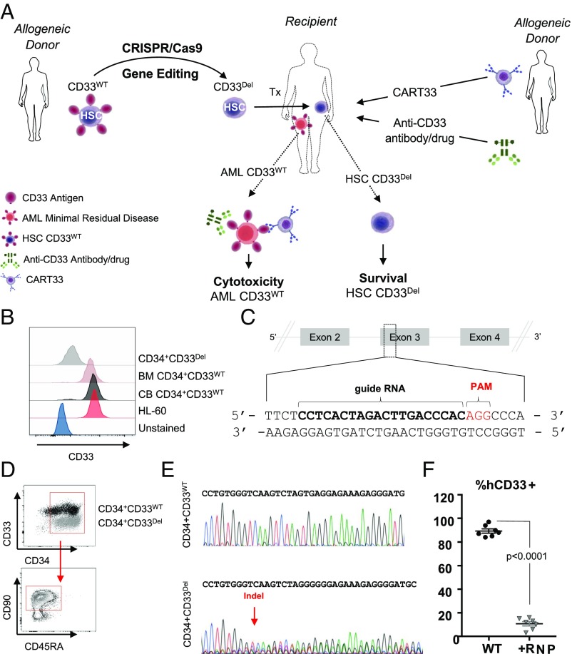
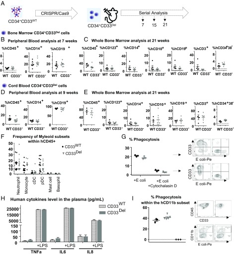
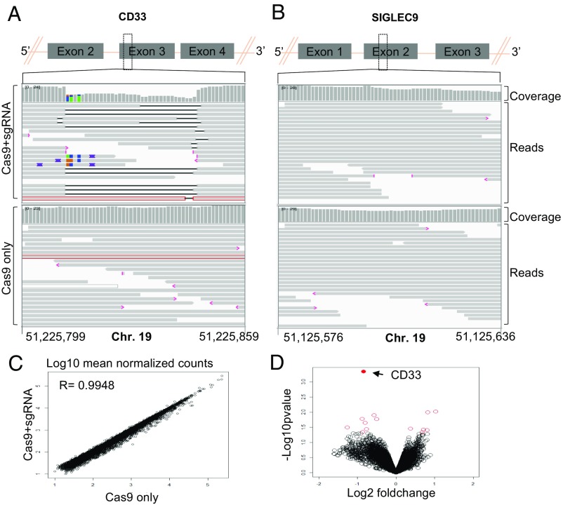
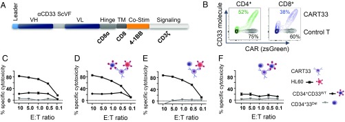
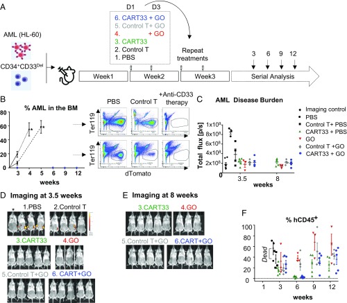
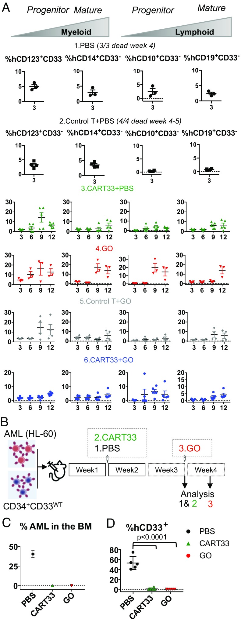
Antigen-directed immunotherapies for acute myeloid leukemia (AML), such as chimeric antigen receptor T cells (CAR-Ts) or antibody-drug conjugates (ADCs), are associated with severe toxicities due to the lack of unique targetable antigens that can distinguish leukemic cells from normal myeloid cells or myeloid progenitors. Here, we present an approach to treat AML by targeting the lineage-specific myeloid antigen CD33. Our approach combines CD33-targeted CAR-T cells, or the ADC Gemtuzumab Ozogamicin with the transplantation of hematopoietic stem cells that have been engineered to ablate CD33 expression using genomic engineering methods. We show highly efficient genetic ablation of CD33 antigen using CRISPR/Cas9 technology in human stem/progenitor cells (HSPC) and provide evidence that the deletion of CD33 in HSPC doesn't impair their ability to engraft and to repopulate a functional multilineage hematopoietic system in vivo. Whole-genome sequencing and RNA sequencing analysis revealed no detectable off-target mutagenesis and no loss of functional p53 pathways. Using a human AML cell line (HL-60), we modeled a postremission marrow with minimal residual disease and showed that the transplantation of CD33-ablated HSPCs with CD33-targeted immunotherapy leads to leukemia clearance, without myelosuppression, as demonstrated by the engraftment and recovery of multilineage descendants of CD33-ablated HSPCs. Our study thus contributes to the advancement of targeted immunotherapy and could be replicated in other malignancies.

Keywords: CD33; CRISPR/Cas9; acute myeloid leukemia; chimeric antigen receptor; transplantation.


Conflict of interest statement

Conflict of interest statement: This study was funded by a grant from Vor Biopharma and PureTech Health, which has launched a company called Vor Biopharma. Columbia University owns equity in Vor Biopharma and has licensed technology that is the subject of this study to Vor Biopharma. F.B., A.M.A., and S.M. are coinventors on issued and pending patent applications licensed to Vor Biopharma. S.M. has equity ownership and is on the Scientific Advisory Board of Vor Biopharma. A.R. received funding from PureTech Health.

Figures

Fig. 1.
Fig. 1.
(A) Approach: stem cells, either mobilized or CB obtained from a donor, will be genetically manipulated to ablate CD33 expression using gene-editing technology, such as CRISPR/Cas9, and transplanted to relapsed patients eligible for HSCT. Subsequent to transplantation, T cells from an allogeneic donor will be genetically manipulated, using a viral delivery system, to express chimeric antigen receptors targeting CD33 and infused in the recipient. Alternatively, patients can receive ADC (GO) either alone or in combination with CAR-T. (BF) CD33 expression and its ablation in human cells. (B) Expression of CD33 in human AML cell line HL-60, in human primary CD34+CD33WT cells from BM and CB and human primary CD34+CD33Del after CRISPR/Cas9-mediated ablation. (C) Schematic representation of CD33 genomic locus showing exons 2–4 and location and sequence of sgRNA (in bold, PAM in red) targeting CD33. (D) Surface expression of CD33 by flow cytometry after electroporation in CD34+CD33WT cells and CD34+CD33Del. All cells maintain their stem cells phenotype as assessed by CD90 expression. (E) Chromatogram of Sanger sequencing showing a region surrounding the DNA double-strand break site, (Upper) CD34+CD33WT cells and (Lower) CD34+CD33Del. (F) Five to 7 d after electroporation, CD34+ cultured cells show consistent deletion of CD33 compared with control (15 independent donors).
Fig. 2.
Fig. 2.
Deletion of CD33 does not impair engraftment, hematopoietic repopulation, and function in NSGS mice. (A) Schematic of experimental design. (B and C) BM-derived CD34+ cells engraftment and repopulation: (B) Peripheral blood (7 wk) and (C) whole BM (21 wk) posttransplant analyzed for cells of various lineages, as indicated. CD34+CD33Del cells show the same engraftment (CD45+) as control cells as well as comparable percentage of mature myeloid and lymphoid cells. BM CD34+CD33Del cells show comparable percentage of myeloid (progenitor CD123+, mature CD14+), and lymphoid (progenitor CD10+, mature CD19+) T cells (CD3+) and stem cells CD34+38. (D and E) CB-derived CD34+ cell engraftment and repopulation: (D) Peripheral blood (9 wk) and (E) BM (21 wk) posttransplant analyzed for cells of various lineages, as indicated. CD34+CD33Del cells show same engraftment (CD45+) as control cells, as well as comparable percentage of mature myeloid and lymphoid cells. BM CD34+CD33Del cells show comparable percentage of myeloid (progenitor CD123+, mature CD14+) and lymphoid (progenitor CD10+, mature CD19+), T cells (CD3+), and stem cells CD34+38. Data were analyzed using unpaired t test and no significant differences were found in all of the groups examined (P > 0.05). All data are represented as mean ± SEM (two independent experiments, two donors). (FI) In vitro and in vivo functional assays. (F) CD34+CD33Del show comparable development of myeloid lineage than CD34+CD33WT in NSGS mice. Frequencies of neutrophils, monocytes, cDC, pDC, mast cells, and basophils in the BM aspirates of NSGS mice injected with CB CD34+CD33WT or CD34+CD33Del cells. (Control n = 12, CD34+CD33Del n = 13). (G) In vitro E. coli bioparticles phagocytosis assay of in vitro CD33WT or CD33Del differentiated monocytes. CD33Del monocytes show similar phagocytosis capacity (two independent experiments, two donors). (H) Response to LPS-induced Toll-like receptor activation is similar in NSGS mice injected with CD34+CD33WT or CD34+CD33Del cells. Analysis of plasma cytokines level at 0 and 4h30 after intraperitoneal injection of 15 μg LPS (Control n = 12, CD34+CD33Del n = 13). (I) Peritoneal cavity analysis 2 h after intravenous injection of E. coli bioparticles (Control n = 3 CD34+CD33Del n = 5), untreated mice (♦). Mouse and syringe images designed by Freepik and Kiranshastry from Flaticon.
Fig. 3.
Fig. 3.
(A and B) IGV screenshot of genomic region of CD33 (A) and SIGLEC9 (B) genes surrounding the guides in Cas9+sgRNA (Top) and Cas9 only (Bottom) cells as indicated in the left. The gray bars in the coverage track (indicated on right) show the depth of the reads displayed at each locus. Generally, the coverage should be uniform and hence the bar height should be same but deletions results in dip in the height. The reads track shows all of the reads (gray boxes) mapped in this region. The deletions are represented by a solid black line and insertions with purple boxes. Reads with red border are those without a mapped mate. One read in each group was without a mapped mate. Mismatch bases are colored in green, blue, brown, and red for nucleotides A, C, G, and T, respectively. SIGLEC9 genomic region was selected as representative region to show absence of indels at off-target site because (i) it belongs to SIGLEC family with homology to CD33, and (ii) it has the highest homology within 10 bp of the expected cut site compared with any other guide (SI Appendix, Table S2). Chromosome coordinates at the bottom are based on hg38. (C) Scatter plot showing correlation between log10 mean normalized counts, normalized using the DEseq2 method, between CD33 edited cells and control cells. (D) Volcano plot showing log2 fold-change and −log10 P value for genes analyzed using the edgeR method; genes that were significantly differentially expressed (P < 0.05) are shown as red open circles and the CD33 gene is represented by a filled red circle and indicated by a left arrow (four donors).
Fig. 4.
Fig. 4.
CD33 deletion protects CD34+ cells from CART33 cytotoxicity in vitro. (A) Schematic of CART33 construct. (B) Contour plot showing CAR expression in human primary T cells after lentiviral transduction with control (black) or CART33 (green or blue) virus. Percentage transduction in each group is specified next to the plots. CD4+ and CD8+ cells were transduced independently and comixed 1:1 before experiment. (CF) Cytotoxicity assays. (C) CART33 cells or control T were incubated with HL-60 or CD34+CD33WT or CD34+CD33Del and cytotoxicity assessed by flow cytometry. (DF) Triple culture cytotoxicity assay. CART33 cells or control T cells were coincubated with (D) HL-60 and CD34+CD33WT or (E) with HL-60 and CD34+CD33Del or (F) with CD34+CD33WT and CD34+CD33Del cells (four independent experiments).
Fig. 5.
Fig. 5.
Therapy model: CD34+CD33Del cells resist CD33-targeted immunotherapy. (A) Schematic of experimental design: 5 × 10⁵ HL-60 and 5 × 10⁵ CD34+CD33Del were injected in NSGS mice on day 0. One week after, mice were treated with PBS or allogeneic CART33 or control T cells. Three days after a new group received GO only, while allogeneic CART33 and control T cells-injected mice received GO or PBS. Treatment was repeated on week 3. Leukemia progression and CD34+CD33Del engraftment were then monitored by serial BM aspiration. (B) Monitoring of leukemia burden in BM aspirates. Leukemia burden in the whole BM of control groups mice at the time of death are shown with an asterisk (*). Leukemia cells were gated on Ter119dtomato+. (C) Leukemia burden measure via epifluorescence quantification of images shown in D and SI Appendix, Fig. S5C at 3.5 wk, and E and SI Appendix, Fig. S5D at 8 wk. One mouse representative of each treatment is shown in C and E. See SI Appendix, Fig. S5 for full imaging panel. Background was removed with untreated mouse (*Imaging control). (F) CART33 or GO leukemia clearance doesn’t impair engraftment of CD34+CD33Del cells overtime (% hCD45+ cells), as shown by flow cytometry of BM aspirates. CD34+-injected derived human cells were gated on Ter119dtomato, Ly5/H2kdhuman CD45+CART (three independent experiments with GO, two independent experiments with CART33). Mouse and syringe images designed by Freepik and Kiranshastry from Flaticon.
Fig. 6.
Fig. 6.
(A) CD34+CD33Del cells resist CD33-targeted immunotherapy and contribute to myelopoiesis and lymphopoiesis. Left two panels in each condition is monitoring overtime of the repopulation of myeloid progenitors, and Right two panels shows lymphoid progenitors and mature cells in BM aspirates. No significant differences were observed between different treatment groups at all time points analyzed. (BD) CD34+CD33WT cells are sensitive to CD33-targeted immunotherapy. (B) A schematic of experimental design: 5 × 10⁵ CD34+CD33WT alone or in combination with 5 × 10⁵ HL-60 were injected in NSGS mice on day 0. One week after, mice were treated with PBS or allogeneic CART33 cells. Leukemia progression and CD34+CD33WT engraftment were then monitored by bone marrow aspiration at week 3 for CART33. The same day a group of mice was injected with GO and analyzed 4 d after. (C and D) BM aspirates show complete elimination of CD33WT leukemia cells (C) and CD33WT primary cells (D) in mice treated with CART33 or GO compared with PBS alone. Significant difference was observed between CART33 and GO compared with PBS. CD34+ injected derived human cells were gated on Ter119dtomato, Ly5/H2kd human CD45+CART. All data are represented as mean ± SEM (two independent experiments, two donors). Mouse and syringe images designed by Freepik and Kiranshastry from Flaticon.

References

    1. O’Donnell M. R., et al. , Acute myeloid leukemia, version 3.2017, NCCN Clinical Practice Guidelines in Oncology. J. Natl. Compr. Canc. Netw. 15, 926–957 (2017). - PubMed
    1. Rai K. R., et al. , Treatment of acute myelocytic leukemia: A study by cancer and leukemia group B. Blood 58, 1203–1212 (1981). - PubMed
    1. Alcantara M., Tesio M., June C. H., Houot R., CAR T-cells for T-cell malignancies: Challenges in distinguishing between therapeutic, normal, and neoplastic T-cells. Leukemia 32, 2307–2315 (2018). - PMC - PubMed
    1. Morgan R. A., et al. , Case report of a serious adverse event following the administration of T cells transduced with a chimeric antigen receptor recognizing ERBB2. Mol. Ther. 18, 843–851 (2010). - PMC - PubMed
    1. Lamers C. H., et al. , Treatment of metastatic renal cell carcinoma with CAIX CAR-engineered T cells: Clinical evaluation and management of on-target toxicity. Mol. Ther. 21, 904–912 (2013). - PMC - PubMed

Publication types