. 2019 May 28;10(1):2342.
doi: 10.1038/s41467-019-10280-3.

Structure of the SARS-CoV nsp12 polymerase bound to nsp7 and nsp8 co-factors

Affiliations

Structure of the SARS-CoV nsp12 polymerase bound to nsp7 and nsp8 co-factors

Robert N Kirchdoerfer et al. Nat Commun. .

Abstract

Recent history is punctuated by the emergence of highly pathogenic coronaviruses such as SARS- and MERS-CoV into human circulation. Upon infecting host cells, coronaviruses assemble a multi-subunit RNA-synthesis complex of viral non-structural proteins (nsp) responsible for the replication and transcription of the viral genome. Here, we present the 3.1 Å resolution structure of the SARS-CoV nsp12 polymerase bound to its essential co-factors, nsp7 and nsp8, using single particle cryo-electron microscopy. nsp12 possesses an architecture common to all viral polymerases as well as a large N-terminal extension containing a kinase-like fold and is bound by two nsp8 co-factors. This structure illuminates the assembly of the coronavirus core RNA-synthesis machinery, provides key insights into nsp12 polymerase catalysis and fidelity and acts as a template for the design of novel antiviral therapeutics.


Conflict of interest statement

The authors declare no competing interests.

Figures
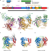
Fig. 1
Fig. 1
Structure of SARS-CoV nsp12 bound to nsp7 and nsp8 co-factors. a Diagram of the SARS-CoV nsp7, nsp8, and nsp12 proteins indicating domains, conserved motifs, and the protein regions observed in the structure (black bars) (Supplementary Fig. 3). b SARS-CoV nsp12 contains a large N-terminal extension composed of the NiRAN domain (dark red) and an interface domain (purple) adjacent to the polymerase domain (orange). nsp12 binds to a heterodimer of nsp7 (blue) and nsp8 (green) as well as to a second subunit of nsp8. ce Comparison of SARS-CoV nsp12 to the polymerase proteins of poliovirus (3OL6.pdb) [10.2210/pdb3OL6/pdb] and dengue virus (4V0R.pdb) [10.2210/pdb4V0R/pdb]. Viral RNA polymerases share an overall structural architecture with fingers (blue), palm (yellow), and thumb domains (red). Both SARS-CoV and dengue virus polymerase proteins contain N-terminal extensions colored green. The SARS-CoV nsp7 and nsp8 cofactors are colored in white and gray, respectively
Fig. 2
Fig. 2
Surface electrostatics and sequence conservation of the nsp7-nsp8-nsp12 complex. a The RNA-binding cleft of SARS-CoV nsp7-nsp8-nsp12 is positively charged while the remaining protein surface carries a negative charge. b Sequence conservation, across the coronavirus family indicates a high level of conservation in the polymerase-active site and nsp7-nsp8 heterodimer-binding site with the binding site of the single nsp8 subunit less conserved. c, d Sequence motifs of the NiRAN domain form a single surface on nsp12 (yellow). Motif CN (blue) and the C-terminal portion of motif BN (pink) are observed in the structure. Amino acids conserved across the Nidovirales order are shown as sticks. Coloring in d is the same as in b
Fig. 3
Fig. 3
Architecture of the nsp12 polymerase-active site. a RNA polymerases possess seven conserved motifs (motifs A–G) involved in template binding (motif G), NTP binding (motif F), and polymerization (motifs A–E). b Superposition of elongation complexes from poliovirus (3OL6.pdb) [10.2210/pdb3OL6/pdb] and norovirus (3H5Y.pdb) [10.2210/pdb3H5Y/pdb] polymerases give the approximate positions of RNA template (red), primer (dark blue), incoming NTP (blue), and bound catalytic metal ions (purple)
Fig. 4
Fig. 4
Comparison of nsp8 conformations. Two conformations of nsp8 are observed in the nsp12-nsp7-nsp8 structure. a A heterodimer of nsp7 (blue) and nsp8 (green) contacts nsp12 (yellow) primarily using surfaces of nsp7. b A second nsp8 subunit also contacts nsp12 directly using a unique conformation of nsp8 amino acids 98–127. The nsp7-nsp8 structures in two conformations for SARS-CoV (2AHM.pdb) [10.2210/pdb2AHM/pdb] (c, d) as well as nsp8 (light green) and two subunits of nsp7 (cyan and purple) for Feline coronavirus (3UB0.pdb) [10.2210/pdb3UB0/pdb] (e) show similar nsp8 conformations as observed in the SARS-CoV nsp7-nsp8 heterodimer bound to nsp12 (a)
Fig. 5
Fig. 5
The nsp7-nsp8 heterodimer stabilizes loops of the fingers domain. a The nsp7-nsp8 heterodimer binds nsp12 on the fingers domain (blue) index finger loop adjacent to the palm (yellow) thumb (red) domains. b In the absence of the nsp7-nsp8 heterodimer, the fingers domain index and ring finger loops as well as several connecting loops in the thumb domain become disordered (delineated by dashed lines). The nsp12 N-terminal extension, nsp7, and nsp8 are shown in black, gray, and white, respectively

References

    1. van der Hoek L. Human coronaviruses: what do they cause? Antivir. Ther. 2007;12:651–658. - PubMed
    1. Centers for Disease Control and Prevention (CDC). Update: severe acute respiratory syndrome--United States, June 11, 2003. MMWR Morb. Mortal Wkly. Rep.52, 550 (2003). - PubMed
    1. Parry J. Breaches of safety regulations are probable cause of recent SARS outbreak, WHO says. BMJ. 2004;328:1222. doi: 10.1136/bmj.328.7442.s122-a. - DOI - PMC - PubMed
    1. Ge XY, et al. Isolation and characterization of a bat SARS-like coronavirus that uses the ACE2 receptor. Nature. 2013;503:535–538. doi: 10.1038/nature12711. - DOI - PMC - PubMed
    1. Ziebuhr J. The coronavirus replicase. Curr. Top. Microbiol. Immunol. 2005;287:57–94. - PMC - PubMed

Publication types

MeSH terms

LinkOut - more resources