. 2019 May 28;9(1):7932.
doi: 10.1038/s41598-019-44333-w.

CGRP signalling inhibits NO production through pannexin-1 channel activation in endothelial cells

Affiliations

CGRP signalling inhibits NO production through pannexin-1 channel activation in endothelial cells

Pablo S Gaete et al. Sci Rep. .

Abstract

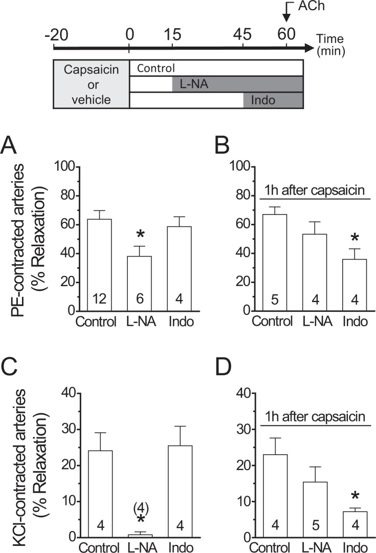
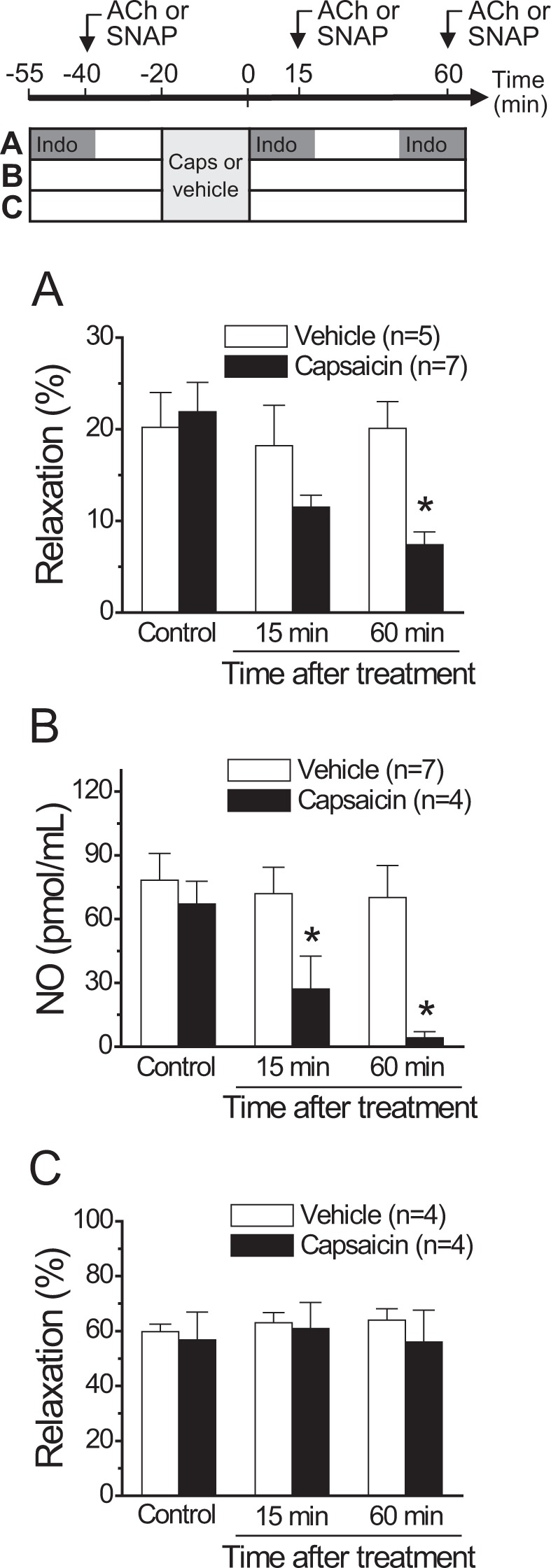
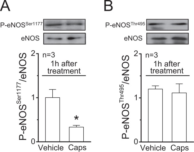
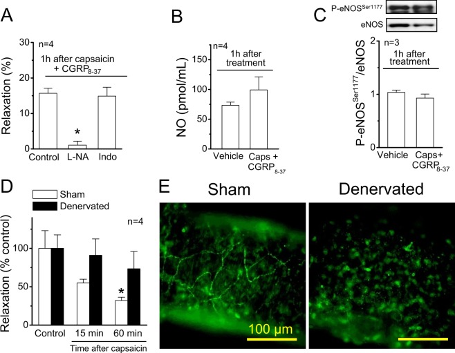
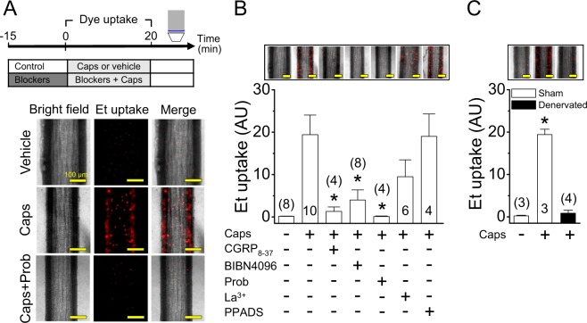
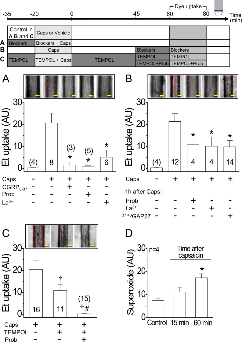
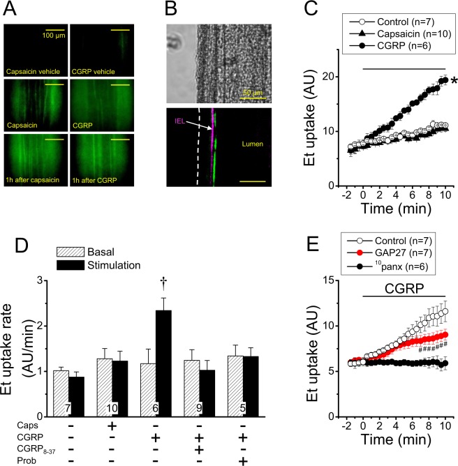
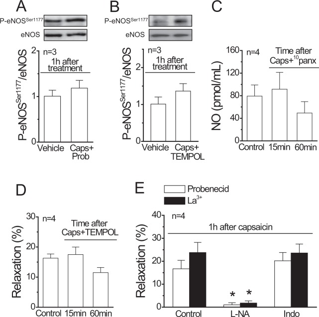
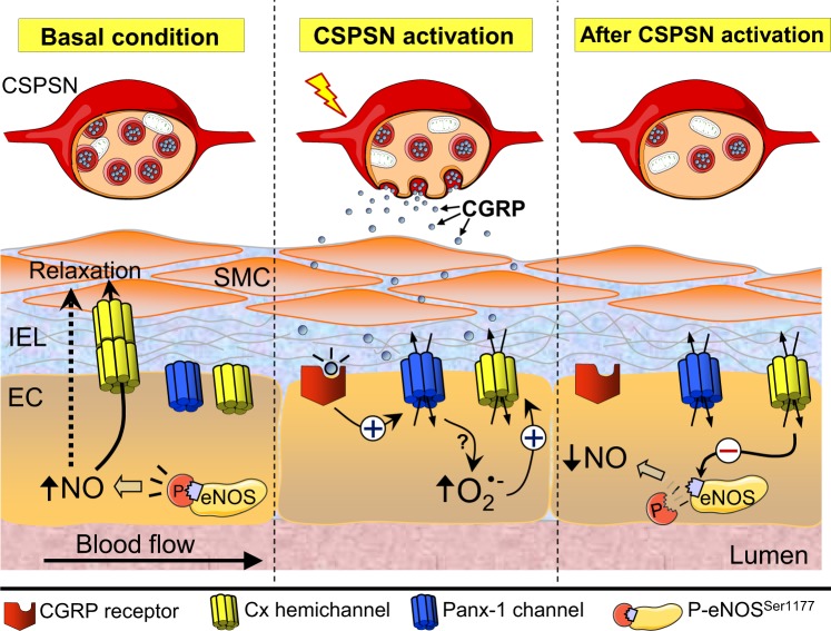
Blood flow distribution relies on precise coordinated control of vasomotor tone of resistance arteries by complex signalling interactions between perivascular nerves and endothelial cells. Sympathetic nerves are vasoconstrictors, whereas endothelium-dependent NO production provides a vasodilator component. In addition, resistance vessels are also innervated by sensory nerves, which are activated during inflammation and cause vasodilation by the release of calcitonin gene-related peptide (CGRP). Inflammation leads to superoxide anion (O2• -) formation and endothelial dysfunction, but the involvement of CGRP in this process has not been evaluated. Here we show a novel mechanistic relation between perivascular sensory nerve-derived CGRP and the development of endothelial dysfunction. CGRP receptor stimulation leads to pannexin-1-formed channel opening and the subsequent O2• --dependent connexin-based hemichannel activation in endothelial cells. The prolonged opening of these channels results in a progressive inhibition of NO production. These findings provide new therapeutic targets for the treatment of the inflammation-initiated endothelial dysfunction.


Conflict of interest statement

The authors declare no competing interests.

Figures

Figure 1
Figure 1
Activation of capsaicin-sensitive sensory nerves triggers the inhibition of ACh-induced NO-dependent vasodilation. The experimental protocol of these results is depicted on the top of the figure. (A) Relaxation induced by 100 nM ACh in phenylephrine (PE)-contracted mesenteric arteries in control conditions and after blockade of NO production with 100 µM NG-nitro-L-arginine (L-NA) or prostaglandin formation with 10 µM indomethacin (Indo). (B) NO- and prostaglandin-dependent vasodilator components activated by ACh 1 h after capsaicin treatment (1 µM, 20 min) in PE-contracted arteries. (C) ACh-elicited relaxation of KCl-contracted mesenteric arteries in the absence and presence of L-NA or Indo. (D) L-NA and Indo sensitive vasodilator components evoked by ACh 1 h after capsaicin treatment in KCl-contracted arteries. Note that the capsaicin-triggered NO signalling inhibition was fully compensated by the activation of an indomethacin-sensitive vasodilator component. Numbers inside the bars or in parentheses indicate the n value. Values are means ± SEM. *P < 0.05 vs control by one-way ANOVA plus Dunnett post hoc test.
Figure 2
Figure 2
Time course of the capsaicin-initiated inhibition of the ACh-induced NO-dependent vasodilator component. The experimental protocol of the results shown in A, B and C is depicted on the top of the figure. (A) Relaxation induced by 100 nM ACh in KCl-contracted mesenteric arteries in control conditions and 15 or 60 min after treatment with capsaicin (1 µM) or its vehicle for 20 min. ACh was applied in the presence of 10 µM indomethacin to prevent the interference of prostaglandins in the response. (B) NO production induced by successive ACh (100 nM) stimulations along the time in mesenteric arterial beds treated with capsaicin or the vehicle. (C) Relaxation induced by 300 nM SNAP, a NO donor, in KCl-contracted mesenteric arteries before (control) or 15 or 60 min after treatment with capsaicin or its vehicle. Values are means ± SEM. *P < 0.05 vs control by one-way ANOVA plus Dunnett post hoc test.
Figure 3
Figure 3
Stimulation of perivascular capsaicin-sensitive sensory nerves leads to a reduction of eNOS phosphorylation at serine 1177. (A,B) Representative Western blots and densitometric analysis of eNOS phosphorylation at serine 1177 (P-eNOSSer1177, panel (A)) and at threonine 495 (P-eNOSThr495, panel (B)) 1 h after the treatment of mesenteric arteries with 1 µM capsaicin (Caps) or the vehicle for 20 min. Changes in eNOS phosphorylation are expressed as the ratio of phosphorylated protein over total protein. Values are means ± SEM. *P < 0.05 vs vehicle by Student’s unpaired t-test.
Figure 4
Figure 4
The capsaicin-initiated NO signalling inhibition is mediated by CGRP release from perivascular sensory nerves. (A) Relaxation induced by 100 nM ACh in KCl-contracted mesenteric arteries 1 h after capsaicin application (1 µM, 20 min) in the presence of 300 nM CGRP8–37. Vasodilator responses were evaluated in control conditions and after the treatment with 100 µM NG-nitro-L-arginine (L-NA) or 10 µM indomethacin (Indo). (B) NO production induced by ACh (100 nM) observed in mesenteric arterial beds 1 h after the treatment with capsaicin in the presence of 300 nM CGRP8–37 or the vehicle. (C) Representative Western blots and densitometric analysis of eNOS phosphorylation at serine 1177 (P-eNOSSer1177) in mesenteries treated 1 h before with capsaicin (Caps) in the presence of 300 nM CGRP8–37 or the vehicle of capsaicin. (D) Relaxation induced by 100 nM ACh in KCl-contracted resistance arteries of sham and denervated mesenteries before (control) and 15 or 60 min after the treatment with capsaicin (1 µM, 20 min). ACh was applied in the presence of 10 µM indomethacin to prevent the interference of prostaglandins in the response, as shown in the experimental protocol depicted in Fig. 2. (E) Detection of perivascular sensory nerves through immunofluorescence analysis of CGRP expression. Values are means ± SEM. *P < 0.001 vs control by one-way ANOVA plus Dunnett post hoc test. Scale bars represent 100 µm.
Figure 5
Figure 5
Activation of capsaicin-sensitive sensory nerves leads to CGRP receptor-mediated Panx-1-formed channel opening. (A) Experimental protocol and representative images of ethidium (Et) uptake observed during vehicle or 1 µM capsaicin (Caps) application for 20 min in mesenteric resistance arteries in the absence and presence of 1 mM probenecid (Prob), a Panx-1 blocker. (B) Representative images and densitometric analysis of Et uptake attained during the stimulation for 20 min with the vehicle of capsaicin or capsaicin in the absence and presence of 300 nM CGRP8–37, 10 µM BIBN4096, 1 mM probenecid (Prob), 200 µM La3+ and 10 µM PPADS. (C) Capsaicin-induced Et uptake observed in control (Sham-operated rats) and denervated mesenteric arteries. Changes in ethidium-fluorescence signal are expressed in arbitrary units (AU). Numbers inside the bars or in parentheses indicate the n value. Values are means ± SEM. *P < 0.05 vs caps (B) or vehicle (C) by one-way ANOVA plus Dunnett post hoc test. Scale bars represent 100 µm.
Figure 6
Figure 6
Activation of Panx-1 channels by capsaicin persists along the time and recruits connexin-based hemichannels through a O2• −-dependent pathway. The experimental protocol of the results shown in A, B and C is depicted on the top of the figure. (A) Representative images and densitometric analysis of the ethidium (Et) uptake attained in 20 min. Et uptake was evaluated 1 h after the end of the application of 1 µM capsaicin (Caps, 20 min) in mesenteric resistance arteries. Capsaicin was applied in the absence and presence of 300 nM CGRP8–37, 1 mM probenecid (Prob) or 200 µM La3+. (B) Et uptake observed 1 h after the end of capsaicin application. Et was perfused for 20 min in the presence of 1 mM probenecid (Prob), 200 µM La3+ or 200 µM 37,43GAP27. (C) Et uptake observed 1 h after the end of capsaicin application. As shown in panel B, ethidium was perfused for 20 min, but, in this case, in the presence of TEMPOL alone or in combination with probenecid (Prob). (D) O2• − production recorded before or 15 and 60 min after capsaicin treatment (1 µM, 20 min). Changes in ethidium-fluorescence signal and O2• − formation are expressed in arbitrary units (AU). Numbers inside the bars or in parentheses indicate the n value. Values are means ± SEM. *P < 0.05 vs caps (A,B) or control (D) by one-way ANOVA plus Dunnett post hoc test. P < 0.05 vs caps and #P < 0.05 vs caps + TEMPOL by one-way ANOVA plus Newman-Keuls post hoc test. Scale bars represent 100 µm.
Figure 7
Figure 7
The CGRP-mediated increase in ethidium uptake induced by capsaicin is initiated by Panx-1-formed channel opening in endothelial cells. (A) Representative images of the Lucifer Yellow uptake (green) induced by perfusion of 1 µM capsaicin and 100 nM CGRP in mesenteric resistance arteries during the stimulation period (10 min) or 1 h after. (B) Representative images of Lucifer Yellow uptake attained after 20 min stimulation of isolated mesenteric resistance arteries with 1 µM capsaicin. In these experiments, Lucifer Yellow and capsaicin were applied in the bath solution. Note that the Lucifer Yellow fluorescent signal is only observed at the inner side of the internal elastic lamina (IEL), confirming that capsaicin activated the uptake of this dye exclusively in the endothelium. (C) Time course of the ethidium (Et) uptake achieved in control conditions and after the stimulation with 1 µM capsaicin or 100 nM CGRP in primary cultures of mesenteric endothelial cells. The horizontal bar indicates the period of stimulation. (D) Analysis of the Et uptake rate observed during the stimulation with capsaicin (Caps) or CGRP in control conditions and in the presence of 1 µM CGRP8–37 or 1 mM probenecid (Prob). The rate of ethidium uptake was assessed by calculating the slope of the increase in fluorescence intensity along the time in basal conditions and during the stimulation period. (E) Time course of the ethidium uptake induced by CGRP in the absence and presence of a combination of the connexin blocking peptides 37,43GAP27 (200 µM) plus 40GAP27 (200 µM) or the Panx-1 blocking peptide 10panx (60 µM). Note that, after treatment with GAP 27 peptides, the ethidium uptake curve shows two components: an initial increase similar to control that starts to decline after 6 min. Changes in ethidium-fluorescence signal are expressed in arbitrary units (AU). Numbers inside the bars indicate the n value. Values are means ± SEM. *P < 0.05 vs control by one-way ANOVA plus Dunnett post hoc test. P < 0.01 vs basal by Student’s paired t-test. #P < 0.05 vs control by two-way ANOVA plus Fisher´s LSD post hoc test.
Figure 8
Figure 8
Participation of Panx-1 channels, connexin hemichannels and O2• − production in the progressive inhibition of the NO-dependent relaxation triggered by capsaicin-sensitive perivascular sensory nerve activation. (A,B) Representative Western blots and densitometric analysis of eNOS phosphorylation at serine 1177 (P-eNOSSer1177) in mesenteries 1 h after the treatment with the vehicle of capsaicin or capsaicin (Caps) in the presence of probenecid (Prob, A) or TEMPOL (B). (C) ACh (100 nM)-induced NO production in mesenteric arterial beds before (control) and 15 or 60 min after capsaicin application in the presence of the Panx-1 blocking peptide 10panx (60 µM). (D) Relaxation induced by 100 nM ACh in KCl-contracted resistance arteries observed in the presence of TEMPOL before (control) and 15 or 60 min after the treatment with capsaicin (1 µM, 20 min). The treatment with TEMPOL started 15 min before the stimulation with ACh in control conditions and was maintained until the end of the experiment. (E) Relaxation induced by 100 nM ACh in KCl-contracted mesenteric arteries 1 h after 1 µM capsaicin application (20 min) in the presence of 1 mM probenecid or 200 µM La3+. The relaxation was evaluated in control conditions and in the presence of 100 µM NG-nitro-L-arginine (L-NA) or 10 µM indomethacin (Indo). Values are means ± SEM. *P < 0.05 vs control by one-way ANOVA plus Dunnett post hoc test.
Figure 9
Figure 9
Schematic model of the inhibition of NO signalling evoked by activation of capsaicin-sensitive perivascular sensory nerves. Control of vasomotor tone in resistance arteries relies on the functional interaction between endothelial cells (EC), smooth muscle cells (SMC) and perivascular nerves, including the capsaicin-sensitive perivascular sensory nerves (CSPSN). Although ECs and SMCs are physically separated by the internal elastic lamina (IEL), vasomotor signals generated by ECs, such as nitric oxide (NO), can reach SMCs either diffusing through the IEL or directly via myoendothelial gap junctions. In resistance arteries, phosphorylation of eNOS at serine 1177 (P-eNOSSer1177) contributes to both the basal control of vasomotor tone and the response induced by endothelium-dependent vasodilators. CGRP release during CSPSN stimulation activates CGRP receptors in ECs, which leads to the opening of pannexin-1 (Panx-1)-formed channels and the further O2• − formation. In turn, the pannexin-1 channel-triggered increase in O2• − causes a long lasting activation of connexin (Cx)-formed hemichannels that provokes a reduction in eNOS phosphorylation at serine 1177, with the consequent decrease in NO production.

References

    1. Lillo, M. A., Pérez, F. R., Puebla, M., Gaete, P. S. & Figueroa, X. F. In The Cardiovascular System - Physiology, Diagnostics and Clinical Implications (ed. David Gaze) (Intech, 2012).
    1. Vanhoutte PM, Mombouli JV. Vascular endothelium: vasoactive mediators. Prog Cardiovasc Dis. 1996;39:229–238. doi: 10.1016/S0033-0620(96)80003-X. - DOI - PubMed
    1. Eichler I, et al. Selective blockade of endothelial Ca2+-activated small- and intermediate-conductance K+-channels suppresses EDHF-mediated vasodilation. Br J Pharmacol. 2003;138:594–601. doi: 10.1038/sj.bjp.0705075. - DOI - PMC - PubMed
    1. Ungvari Z, Csiszar A, Koller A. Increases in endothelial Ca(2+) activate K(Ca) channels and elicit EDHF-type arteriolar dilation via gap junctions. Am J Physiol Heart Circ Physiol. 2002;282:H1760–1767. doi: 10.1152/ajpheart.00676.2001. - DOI - PubMed
    1. Forstermann U, Hertting G, Neufang B. The role of endothelial and non-endothelial prostaglandins in the relaxation of isolated blood vessels of the rabbit induced by acetylcholine and bradykinin. Br J Pharmacol. 1986;87:521–532. doi: 10.1111/j.1476-5381.1986.tb10194.x. - DOI - PMC - PubMed

Publication types