

Enhanced osteogenic proliferation and differentiation of human adipose-derived stem cells on a porous n-HA/PGS-M composite scaffold
- PMID: 31138861
- PMCID: PMC6538636
- DOI: 10.1038/s41598-019-44478-8
Enhanced osteogenic proliferation and differentiation of human adipose-derived stem cells on a porous n-HA/PGS-M composite scaffold
Abstract
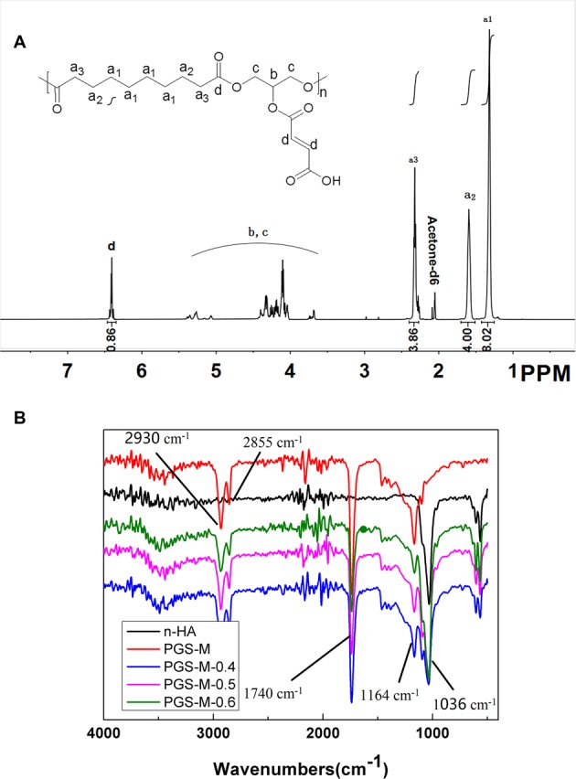
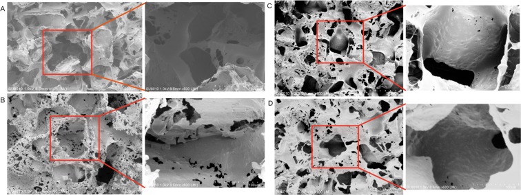
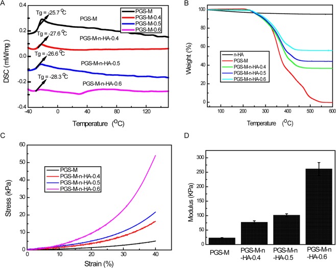
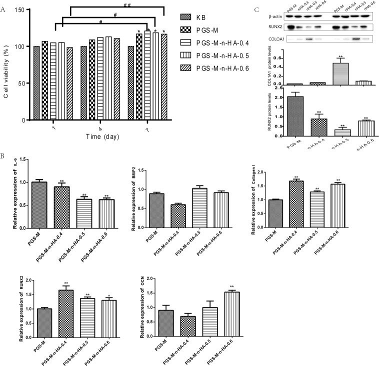
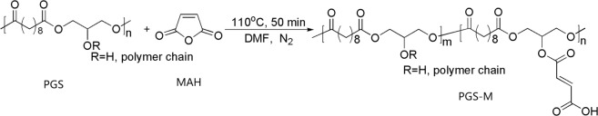
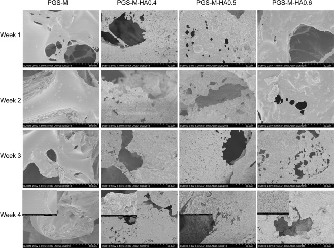
This study explored the applicability, cellular efficacy, and osteogenic activities of porous nano-hydroxyapatite/Poly (glycerol sebacate)-grafted maleic anhydride (n-HA/PGS-g-M) composite scaffolds. Nuclear magnetic resonance (NMR) analyses indicated that approximately 43% of the hydroxide radicals in PGS were displaced by maleic anhydride. Resonance bands at 1036 cm-1 occurred in scaffolds containing nHA powders, and peak areas increased when n-HA weight increased in PGS-M-n-HA-0.4, PGS-M-n-HA-0.5, and PGS-M-n-HA-0.6 scaffolds. The n-HA/PGS-g-M composite scaffolds exhibited porous microstructure with average pore size of 150-300 µm in scanning electron microscopy (SEM) analysis. Differential scanning calorimetry (DSC) identified the glass transition temperature (Tg) as -25-30 °C, indicative of quality resilience. The modulus of compressibility increased when n-HA content increased. Interestingly, viability of human adipose-derived stem cells (hADSCs) in vitro and expression of the osteogenic related genes RUNX2, OCN, and COL1A1 was enhanced in the n-HA/PGS-g-M composite scaffolds compared to those factors observed in PGS-g-M scaffolds. Finally, simulated body fluid (SBF) tests indicated more apatite deposits on the surface of n-HA/PGS-g-M scaffolds compared to PGS-g-M scaffolds. Overall, porous n-HA/PGS-g-M composite scaffolds possessed acceptable biocompatibility and mechanical properties, and they stimulated hADSC cell proliferation and differentiation. Given these qualities, the composite scaffolds have potential applications in bone tissue engineering.
Conflict of interest statement
The authors declare no competing interests.
Figures
Similar articles
-
A poly(glycerol sebacate)-coated mesoporous bioactive glass scaffold with adjustable mechanical strength, degradation rate, controlled-release and cell behavior for bone tissue engineering.Colloids Surf B Biointerfaces. 2015 Jul 1;131:1-11. doi: 10.1016/j.colsurfb.2015.04.031. Epub 2015 Apr 20. Colloids Surf B Biointerfaces. 2015. PMID: 25935647
-
PGS/HAp Microporous Composite Scaffold Obtained in the TIPS-TCL-SL Method: An Innovation for Bone Tissue Engineering.Int J Mol Sci. 2021 Aug 10;22(16):8587. doi: 10.3390/ijms22168587. Int J Mol Sci. 2021. PMID: 34445293 Free PMC article.
-
Synergistic interaction of platelet derived growth factor (PDGF) with the surface of PLLA/Col/HA and PLLA/HA scaffolds produces rapid osteogenic differentiation.Colloids Surf B Biointerfaces. 2016 Mar 1;139:68-78. doi: 10.1016/j.colsurfb.2015.11.053. Epub 2015 Nov 28. Colloids Surf B Biointerfaces. 2016. PMID: 26700235
-
A Review: Optimization for Poly(glycerol sebacate) and Fabrication Techniques for Its Centered Scaffolds.Macromol Biosci. 2021 Sep;21(9):e2100022. doi: 10.1002/mabi.202100022. Epub 2021 Jun 12. Macromol Biosci. 2021. PMID: 34117837 Review.
-
Brief review on poly(glycerol sebacate) as an emerging polyester in biomedical application: Structure, properties and modifications.Polim Med. 2021 Jan-Jun;51(1):43-50. doi: 10.17219/pim/139585. Polim Med. 2021. PMID: 34327876 Review.
Cited by
-
Mesenchymal Stem Cells in Soft Tissue Regenerative Medicine: A Comprehensive Review.Medicina (Kaunas). 2023 Aug 10;59(8):1449. doi: 10.3390/medicina59081449. Medicina (Kaunas). 2023. PMID: 37629738 Free PMC article. Review.
-
Engineering Triphasic Nanocomposite Coatings on Pretreated Mg Substrates for Biomedical Applications.ACS Appl Mater Interfaces. 2024 Oct 9;16(40):54716-54730. doi: 10.1021/acsami.4c13811. Epub 2024 Sep 29. ACS Appl Mater Interfaces. 2024. PMID: 39344064 Free PMC article.
-
Poly(Glycerol Sebacate) in Biomedical Applications-A Review of the Recent Literature.Adv Healthc Mater. 2021 May;10(9):e2002026. doi: 10.1002/adhm.202002026. Epub 2021 Mar 17. Adv Healthc Mater. 2021. PMID: 33733604 Free PMC article. Review.
-
In Vitro Models of Bone Marrow Remodelling and Immune Dysfunction in Space: Present State and Future Directions.Biomedicines. 2022 Mar 24;10(4):766. doi: 10.3390/biomedicines10040766. Biomedicines. 2022. PMID: 35453515 Free PMC article. Review.
-
Translating Material Science into Bone Regenerative Medicine Applications: State-of-The Art Methods and Protocols.Int J Mol Sci. 2022 Aug 22;23(16):9493. doi: 10.3390/ijms23169493. Int J Mol Sci. 2022. PMID: 36012749 Free PMC article. Review.
References
-
- Betz RR. Limitations of autograft and allograft: new synthetic solutions. Orthopedics. 2002;25:561–570. - PubMed
-
- Omar O, Dahlin A, Gasser A, Dahlin C. Tissue dynamics and regenerative outcome in two resorbable non‐cross‐linked collagen membranes for guided bone regeneration: A preclinical molecular and histological study in vivo. Clinical oral implants research. 2018;29:7–19. doi: 10.1111/clr.13032. - DOI - PubMed
MeSH terms
Substances
LinkOut - more resources
Full Text Sources
Other Literature Sources
Miscellaneous
