. 2019 May 8:10:293.
doi: 10.3389/fendo.2019.00293. eCollection 2019.

The Effect of Wnt Pathway Modulators on Human iPSC-Derived Pancreatic Beta Cell Maturation

Affiliations

The Effect of Wnt Pathway Modulators on Human iPSC-Derived Pancreatic Beta Cell Maturation

Heidrun Vethe et al. Front Endocrinol (Lausanne). .

Abstract

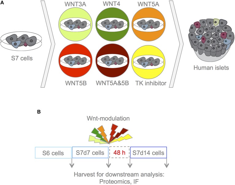
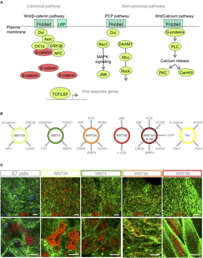
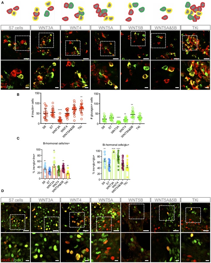
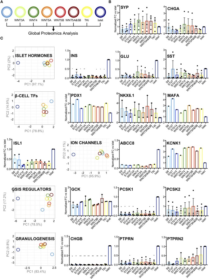
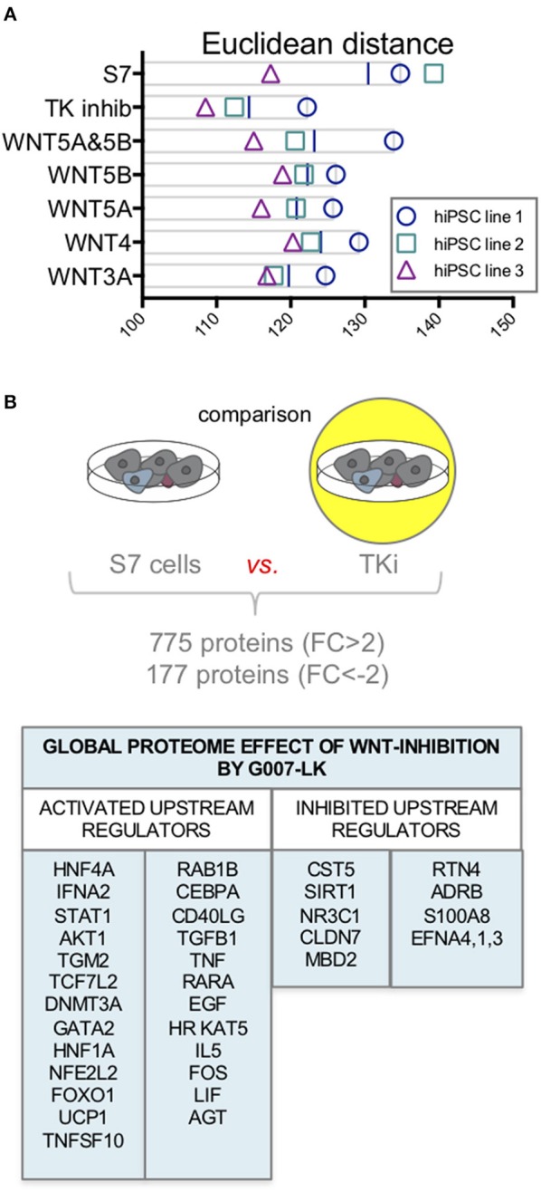
Current published protocols for targeted differentiation of human stem cells toward pancreatic β-cells fail to deliver sufficiently mature cells with functional properties comparable to human islet β-cells. We aimed to assess whether Wnt-modulation could promote the final protocol stages of β-cell maturation, building our hypothesis on our previous findings of Wnt activation in immature hiPSC-derived stage 7 (S7) cells compared to adult human islets and with recent data reporting a link between Wnt/PCP and in vitro β-cell maturation. In this study, we stimulated canonical and non-canonical Wnt signaling in hiPSC-derived S7 cells using syntetic proteins including WNT3A, WNT4, WNT5A and WNT5B, and we inhibited endogenous Wnt signaling with the Tankyrase inhibitor G007-LK (TKi). Whereas neither canonical nor non-canonical Wnt stimulation alone was able to mature hiPSC-derived S7 cells, WNT-inhibition with TKi increased the fraction of monohormonal cells and global proteomics of TKi-treated S7 cells showed a proteomic signature more similar to adult human islets, suggesting that inhibition of endogenous Wnt contributes toward final β-cell maturation.

Keywords: TMT11-plex; Wnt signaling pathway; adult human islets; human induced pluripotent stem cell; in vitro maturation; proteomics; tankyrase inhibition; β-like cells.


Figures

Figure 1
Figure 1
Experimental design. (A) With this experimental set-up we aimed to assess whether Wnt-modulation could drive maturation of S7 cells toward a phenotype that closer resembles that of β-cells as found in adult human islets. To assess the effects of Wnt-modulation of S7 cells, the Wnt-modulated cells were compared to un-stimulated S7 cells as well as to adult human islets. (B) S7d7 cell cultures were treated for 4 h with either WNT3A (light green), WNT4 (green), WNT5A (orange), WNT5B (red), a combination of WNT5A&5B (dark red) for stimulation of the canonical or non-canonical Wnt pathways, or TKi (yellow) to block endogenous Wnt signaling in S7d7 cell cultures. The Wnt-modulated S7 cells were maintained in differentiation culture for 48 h prior to harvest for downstream analysis [including proteomics analysis and immunofluorescence (IF)].
Figure 2
Figure 2
S7-cells show heterogeneous expression of downstream targets 48 h after Wnt-modulator treatment. (A) Schematic overview of key proteins of the canonical and non-canonical Wnt signaling pathways, in brief, Wnt ligand binds to its receptor Frizzled and co-receptors LRP5/6, receptor tyrosine kinase, (or ROR2) and transmits the signal via disheveled (Dvl) into the cytoplasm to activate the canonical Wnt pathway, or functions through non-canonical planar cell polarity (PCP) and Wnt/Ca2+. In the canonical Wnt signaling pathway, β-catenin accumulates in the cytoplasm and translocate to the nucleus to act as a transcription coactivator for the TCF/LEF transcription factor family. Without Wnt, β-catenin is degraded by the destruction complex, composed of Axin, adenomatosis polyposis coli (APC), glycogen synthase kinase 3β (GSK3β) and casein kinase 1α (CK1α). The non-canonical Wnt/PCP pathway is thought to use Ryk or ROR2 for activation; Dvl is recruited to form a complex with disheveled-associated activator of morphogenesis 1 (DAAM1). DAAM1 activates Rho that again activates Rho-associated kinase (ROCK). Dvl can also form a complex with Rac1 to activate JNK via the MAPK pathway. In the Wnt/Ca2+ pathway, binding of Wnt to Frizzled activates a trimetic G-protein leading to activation of PLC to cleave PIP2 to form DAG and IP3. IP3 binds to its receptor on the endoplasmic reticulum and calcium is released. Increased concentrations of calcium and DAG can again activate PKC and CaMKII. (B) A selection of interaction partners of the selected Wnt-ligands (–33). (C) IF of β-catenin, BMP4, ROR2 and c-JUN in S7 cells, WNT3A, WNT4, WNT5A, WNT5B treated S7 cells, respectively. Scale bar 50 μm.
Figure 3
Figure 3
Wnt-modulation influences the distribution of mono-hormonal and bi-hormonal S7 cells. (A) Representative schematic drawing and IF analysis for insulin+ (red), glucagon+ (green) cells and insulin+/glucagon+ cells (yellow) of the respective S7 cell populations. (B) Calculations of insulin+ (red, left) and glucagon+ (green, right) cells were done as described in Methods. The y-axis shows the number of insulin+ and glucagon+ cells, respectively. The figure shows standard error of the mean (SEM) values for each of the columns in bar charts. **P < 0.006, ****P < 0.0001 vs. S7 cells with two-tailed t-test. No significant comparisons show no stars. The number of insulin+ cells and glucagon+ cells were normalized to total cell count (dapi+ cells) see Supplementary Figure 2. (C) Overlay of insulin+ and glucagon+ were used to count bi-hormonal cells, in which bi-hormonal (ins+glu+) cells were calculated as a fraction of insulin+ (red bar chart) and glucagon+ (green bar chart) cells, respectively. The figure shows standard error of the mean (SEM) values for each of the columns in bar charts. **P < 0.006, ****P < 0.0001 vs. S7 cells with two-tailed, type two t-test. No significant comparisons show no stars. (D) IF analysis of NKX6.1+ (red), PDX1+ (green) cells and NKX6.1+/PDX1+ cells (yellow). Scale bar upper panel: 25 μm, lower panel: 7.5 μm.
Figure 4
Figure 4
Pancreatic endocrine markers. (A) Schematic drawing of the color code for the different samples analyzed. (B) Protein abundance of pan-endocrine markers Synaptophysin (SYP) and Chromogranin (CHGA) in S6, S7d7, S7d14, Wnt-modualted S7 cells and adult human islets. The y-axis shows normalized fold change vs. islets for each protein. The data shown is normalized protein levels from all cell lines (n = 3, when detected) and is shown as mean with SEM. (C) PC-plots for hormones (INS, GCG, SST), transcription factors (PDX1, NKX6.1, MAFA, and ISL1), ion channels (ABCC8, KCNK1), GSIS regulators (GCK, PCSK1, PCSK2, LDHA, HK1, HK2, ALDOB) and proteins involved in granulogenesis (CHGA, CHGB, PTPRN, PTPRN2). Unit variance scaling is applied to rows. Singular value decomposition(SVD) with imputation is used to calculate principal components. X and y-axis show principal component 1 and principal component 2 that explain the % of the total variance, respectively. N = 8 data points. Also included are charts showing normalized fold change (y-axis) for each protein vs. islets. The data shown is normalized protein levels from all cell lines (n = 3, when detected) and is shown as mean with SEM. *P < 0.05, **P < 0.006 as vs. islets (normalized abundance level of adult human islets as reference) t-test. No significant comparisons show no stars.
Figure 5
Figure 5
Proteome changes in response to Wnt-modulation of S7 cells. (A) Euclidean distance on log2 scale between S7 cells and the different Wnt-modulated S7 cell conditions and adult human islets. Of the three cell lines (n = 3, biological triplicates), TKi had the lowest distance measured between TKi-treated S7 cells vs. adult human islets. (B) Using quantitative proteomics data from one of the cell lines we examined the global proteome effect of endogenous Wnt inhibition by TKi on S7 cells resulting in increased abundance of 755 proteins (FC > 2) and decreased abundance of 177 proteins (FC < −2). The IPA software predicted upstream regulators protein signature based on these differentially regulated proteins.

References

    1. Rezania A, Bruin JE, Arora P, Rubin A, Batushansky I, Asadi A, et al. . Reversal of diabetes with insulin-producing cells derived in vitro from human pluripotent stem cells. Nat Biotechnol. (2014) 32:1121–33. 10.1038/nbt.3033 - DOI - PubMed
    1. Pagliuca FW, Millman JR, Gurtler M, Segel M, Van Dervort A, Ryu JH, et al. . Generation of functional human pancreatic beta cells in vitro. Cell. (2014) 159:428–39. 10.1016/j.cell.2014.09.040 - DOI - PMC - PubMed
    1. Russ HA, Parent AV, Ringler JJ, Hennings TG, Nair GG, Shveygert M, et al. . Controlled induction of human pancreatic progenitors produces functional beta-like cells in vitro. EMBO J. (2015) 34:1759–72. 10.15252/embj.201591058 - DOI - PMC - PubMed
    1. Johnson JD. The quest to make fully functional human pancreatic beta cells from embryonic stem cells: climbing a mountain in the clouds. Diabetologia. (2016) 59:2047–57. 10.1007/s00125-016-4059-4 - DOI - PubMed
    1. Blum B, Hrvatin SS, Schuetz C, Bonal C, Rezania A, Melton DA. Functional beta-cell maturation is marked by an increased glucose threshold and by expression of urocortin 3. Nat Biotechnol. (2012) 30:261–4. 10.1038/nbt.2141 - DOI - PMC - PubMed