Review
. 2019 Apr 15;9(2):110-126.
eCollection 2019.

Update on imaging-based diagnosis of acute renal allograft rejection

Affiliations
Review

Update on imaging-based diagnosis of acute renal allograft rejection

Richard Köhnke et al. Am J Nucl Med Mol Imaging. .

Abstract

Kidney transplantation is the preferred treatment for patients with end-stage renal disease. Despite effective immunosuppressants, acute allograft rejections pose a major threat to graft survival. In early stages, acute rejections are still potentially reversible, and early detection is crucial to initiate the necessary treatment options and to prevent further graft dysfunction or even loss of the complete graft. Currently, invasive core needle biopsy is the reference standard to diagnose acute rejection. However, biopsies carry the risk of graft injuries and cannot be immediately performed on patients receiving anticoagulation drugs. Therefore, non-invasive assessment of the whole organ for specific and rapid detection of acute allograft rejection is desirable. We herein provide a review summarizing current imaging-based approaches for non-invasive diagnosis of acute renal allograft rejection.

Keywords: Acute allograft rejection; MRI; PET; SPECT; imaging; kidney transplantation; noninvasive; ultrasound.


Conflict of interest statement

None.

Figures

Figure 1
Figure 1
Representative ultrasound images of an allogeneically transplanted (aTX) rat kidney (graft) and its native control kidney (native) on day four post surgery (POD4). Depicted are examples of transversal images taken before (pre-CM) and 15 minutes after (post-CM) tail vein injection of anti-CD3-antibody labeled microbubbles. POD: postoperative day, CM: contrast media/microbubbles conjugated to anti-CD3 antibody.
Figure 2
Figure 2
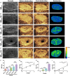
Qualitative and quantitative analysis of C4d deposition in the kidney. Control microbubbles (MBCon) and C4d targeted microbubbles (MBC4d) were injected into recipients and targeted ultrasound (US) imaging was obtained via a destructive-replenishment approach. To ensure the clearance of MBs in circulation, the interval between applications of MBCon and MBC4d was set to 30 min. A-E. Representative targeted US images of the MBcon group (n = 20), MBC4d-G0 group (SY grafts 3 d after transplantation; n = 5), MBC4d-G1 group (6 h after transplantation; n = 5), MBC4d-G2 group (1 d after transplantation; n = 5), and MBC4d-G3 group (3 d after transplantation; n = 5). F. The normalized intensity differences (NIDs) of different C4d grades (n = 20 in the MBcon group; n = 5 in each of other groups). G. Correlation analysis between NID and C4d grades (n = 5/group). H. The integrated optical density (IOD) of different C4d grades (n = 5/group). I. The correlation between IOD and C4d grades (n = 5/group). *P < 0.05; ***P < 0.001. This image has been taken with permission from the paper [21].
Figure 3
Figure 3
In vivo ASL (A) and glucoCEST (B) MRI showing representative ASL and glucoCEST MTRasym maps of an allogeneically transplanted rat on day four post surgery. Decreased perfusion and increased glucose accumulation is detected especially in the cortex of the allograft on the right side (L) compared to the healthy right contralateral kidney on the left side (R).
Figure 4
Figure 4
Representative PET-images of dynamic whole-body acquisitions of a allogeneically transplanted rat (postoperative day four, (A) after tail vein injection of 30 MBq 18F-FDG (maximum a posterior projection, 180 min p.i.) and (B) after tail vein injection of 30·106 18F-FDG-labeled T cells (maximum-intensity projection, 50-70 min p.i.). While the allografts undergoing rejection show distinct enhancement of 18F-FDG (yellow circles) the native control kidneys without rejection do not (green circles).
Figure 5
Figure 5
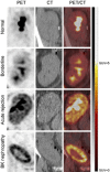
Representative 18F-FDG PET/CT imaging in kidney transplant recipients with suspected acute rejection. PET (left column), CT (middle column), and combined PET/CT images taken after administration of 18F-FDG are shown for kidney transplant recipients with biopsies showing normal histology, borderline changes, acute rejection or polyomavirus BK nephropathy. The arbitrary scale of SUVs (from 0 to 5) is illustrated on the right side. 18F-FDG, fluorodeoxyglucose F18; CT, computed tomography; PET, positron emission tomography; SUV, standard uptake value. This image has been reprinted with permission from the paper [83].

References

    1. Wolfe RA, Ashby VB, Milford EL, Ojo AO, Ettenger RE, Agodoa LY, Held PJ, Port FK. Comparison of mortality in all patients on dialysis, patients on dialysis awaiting transplantation and recipients of a first cadaveric transplant. N Engl J Med. 1999;341:1725–1730. - PubMed
    1. Tabriziani H, Lipkowitz MS, Vuong N. Chronic kidney disease, kidney transplantation and oxidative stress: a new look to successful kidney transplantation. Clin Kidney J. 2017;11:130–135. - PMC - PubMed
    1. Elbadri A, Traynor C, Veitch JT, O’Kelly P, Magee C, Denton M, O’Sheaghdha C, Conlon PJ. Factors affecting eGFR 5-year post-deceased donor renal transplant: analysis and predictive model. Ren Fail. 2015;37:417–423. - PubMed
    1. Jalalzadeh M, Mousavinasab N, Peyrovi S, Ghadiani MH. The impact of acute rejection in kidney transplantation on long-term allograft and patient outcome. Nephrourol Mon. 2015;7:e24439. - PMC - PubMed
    1. Hanssen O, Erpicum P, Lovinfosse P, Meunier P, Weekers L, Tshibanda L, Krzesinski JM, Hustinx R, Jouret F. Non-invasive approaches in the diagnosis of acute rejection in kidney transplant recipients. Part I. in vivo imaging methods. Clin Kidney J. 2017;10:97–105. - PMC - PubMed

LinkOut - more resources