Corpus callosum axon diameter relates to cognitive impairment in multiple sclerosis
- PMID: 31139686
- PMCID: PMC6529828
- DOI: 10.1002/acn3.760
Corpus callosum axon diameter relates to cognitive impairment in multiple sclerosis
Abstract
Objective: To evaluate alterations in apparent axon diameter and axon density obtained by high-gradient diffusion MRI in the corpus callosum of MS patients and the relationship of these advanced diffusion MRI metrics to neurologic disability and cognitive impairment in MS.
Methods: Thirty people with MS (23 relapsing-remitting MS [RRMS], 7 progressive MS [PMS]) and 23 healthy controls were scanned on a human 3-tesla (3T) MRI scanner equipped with 300 mT/m maximum gradient strength using a comprehensive multishell diffusion MRI protocol. Data were fitted to a three-compartment geometric model of white matter to estimate apparent axon diameter and axon density in the midline corpus callosum. Neurologic disability and cognitive function were measured using the Expanded Disability Status Scale (EDSS), Multiple Sclerosis Functional Composite (MSFC), and Minimal Assessment of Cognitive Function in MS battery.
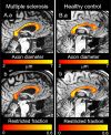
Results: Apparent axon diameter was significantly larger and axon density reduced in the normal-appearing corpus callosum (NACC) of MS patients compared to healthy controls, with similar trends seen in PMS compared to RRMS. Larger apparent axon diameter in the NACC of MS patients correlated with greater disability as measured by the EDSS (r = 0.555, P = 0.007) and poorer performance on the Symbol Digits Modalities Test (r = -0.593, P = 0.008) and Brief Visuospatial Memory Test-Revised (r = -0.632, P < 0.01), tests of interhemispheric processing speed and new learning and memory, respectively.
Interpretation: Apparent axon diameter in the corpus callosum obtained from high-gradient diffusion MRI is a potential imaging biomarker that may be used to understand the development and progression of cognitive impairment in MS.
Conflict of interest statement
None declared.
Figures

References
-
- Rao SM, Leo GJ, Bernardin L, Unverzagt F. Cognitive dysfunction in multiple sclerosis. I. Frequency, patterns, and prediction. Neurology 1991;41:685–691. - PubMed
-
- Benedict RH, Zivadinov R. Risk factors for and management of cognitive dysfunction in multiple sclerosis. Nat Rev Neurol 2011;7:332–342. - PubMed
-
- Evangelou N, Konz D, Esiri MM, et al. Regional axonal loss in the corpus callosum correlates with cerebral white matter lesion volume and distribution in multiple sclerosis. Brain 2000;123(Pt 9):1845–1849. - PubMed
-
- Granberg T, Martola J, Bergendal G, et al. Corpus callosum atrophy is strongly associated with cognitive impairment in multiple sclerosis: results of a 17‐year longitudinal study. Mult Scler 2015;21:1151–1158. - PubMed
MeSH terms
Grants and funding
LinkOut - more resources
Full Text Sources
Medical
