

Quantitative mapping of acute and chronic PCL pathology with 3 T MRI: a prospectively enrolled patient cohort
- PMID: 31139976
- PMCID: PMC6538732
- DOI: 10.1186/s40634-019-0188-2
Quantitative mapping of acute and chronic PCL pathology with 3 T MRI: a prospectively enrolled patient cohort
Abstract
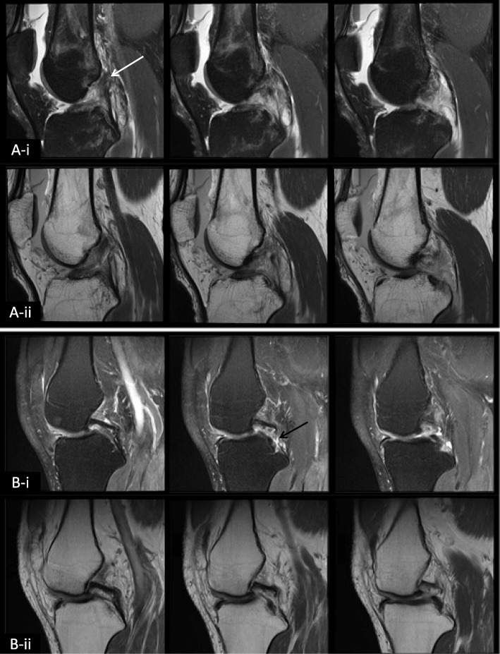
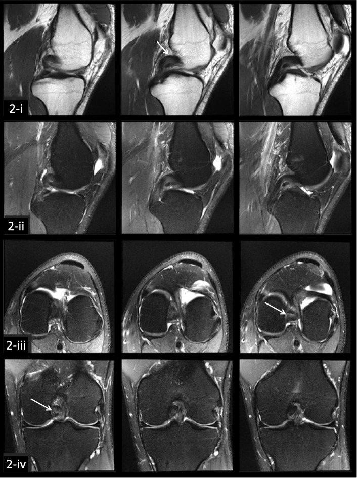
Background: The diagnosis of incomplete acute and chronic posterior cruciate ligament (PCL) tears can be challenging with conventional magnetic resonance (MR) imaging, particularly for injuries in which the ligament appears continuous as occurs with chronic PCL tears that have scarred in continuity. Quantitative mapping from MR imaging may provide additional useful diagnostic information in these cases. The purpose of this study was to assess the feasibility of quantifying transverse relaxation time (T2) mapping values at 3 Tesla (T) in a prospectively enrolled patient cohort with chronic PCL tears.
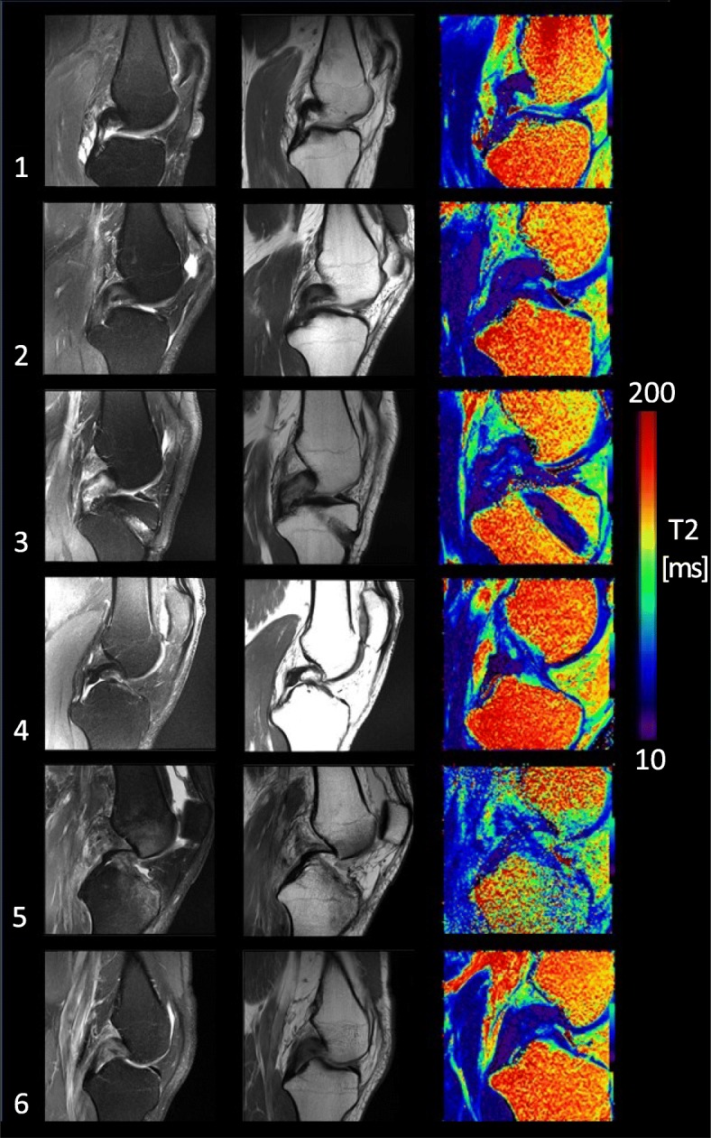
Methods: Twelve subjects with acute or chronic functionally torn PCL, confirmed on clinical exam and posterior knee stress radiographs (with 8 mm or more of increased posterior tibial translation), were enrolled prospectively over a span of 4 years (age: 28-52 years, injury occurred 2 weeks to 15 years prior). Unilateral knee MR images were acquired at 3 T, including a multi-echo spin-echo T2 mapping scan in the sagittal plane. For the six subjects with a continuous PCL on MR imaging the PCL was manually segmented and divided into proximal, mid and distal thirds. Summary statistics for T2 values in each third of the ligament were compiled.
Results: Across the six patient subjects with a continuous ligament, the mean T2 for the entire PCL was 36 ± 9 ms, with the highest T2 values found in the proximal third (proximal: 41 ms, mid 30 ms, distal 37 ms). The T2 values for the entire PCL and for the proximal third subregion were higher than those recently published for asymptomatic volunteers (entire posterior cruciate ligament: 31 ± 5 ms, proximal: 30 ms, mid: 29 ms, distal: 37 ms) with similar methodology.
Conclusion: Mean T2 values were quantified for acute and chronic PCL tears in this prospectively enrolled patient cohort and were higher than those reported for asymptomatic volunteers. This novel approach of using quantitative mapping to highlight injured areas of the posterior cruciate ligament has potential to provide additional diagnostic information in the challenging case of a suspected posterior cruciate ligament tear which appears continuous, including chronic tears that have scarred in continuity and may appear intact on conventional magnetic resonance imaging.
Keywords: Knee; Magnetic resonance imaging; Posterior cruciate ligament; T2 mapping.
Conflict of interest statement
Jurgen Fripp and Craig Engstrom have no potential conflicts of interest. The remainder of the authors have institutional disclosures related to affiliation with the Steadman Philippon Research Institute which is a 501(c) (3) non-profit institution supported financially by private donations and corporate support from the following entities: Smith & Nephew Endoscopy, Arthrex, Siemens Medical Solutions, USA, Ossur Americas, Vail Health Hospital. Additionally, Charles Ho serves on the Steadman Philippon Research Institute Research Advisory Committee and is a consultant for Rotation Medical, not related to this manuscript or research and Robert LaPrade serves on the Steadman Philippon Research Institute Research Advisory Committee and is a consultant for, and receives royalties from, Arthrex, Ossur, and Smith & Nephew.
None of these disclosures are directly related to this research.
Figures
References
-
- Biercevicz AM, Miranda DL, Machan JT, Murray MM, Fleming BC. In situ, noninvasive, T2*-weighted MRI-derived parameters predict ex vivo structural properties of an anterior cruciate ligament reconstruction or bioenhanced primary repair in a porcine model. Am J Sports Med. 2013;41(3):560–566. doi: 10.1177/0363546512472978. - DOI - PMC - PubMed
-
- Chou MC, Tsai PH, Huang GS, Lee HS, Lee CH, Lin MH, Lin CY, Chung HW. Correlation between the MR T2 value at 4.7 T and relative water content in articular cartilage in experimental osteoarthritis induced by ACL transection. Osteoarthr Cartil. 2009;17(4):441–447. doi: 10.1016/j.joca.2008.09.009. - DOI - PubMed
LinkOut - more resources
Full Text Sources
