. 2019 Aug;29(4):195-207.
doi: 10.1089/nat.2019.0784. Epub 2019 May 28.

Formulation of Biocompatible Targeted ECO/siRNA Nanoparticles with Long-Term Stability for Clinical Translation of RNAi

Affiliations

Formulation of Biocompatible Targeted ECO/siRNA Nanoparticles with Long-Term Stability for Clinical Translation of RNAi

Nadia R Ayat et al. Nucleic Acid Ther. 2019 Aug.

Abstract

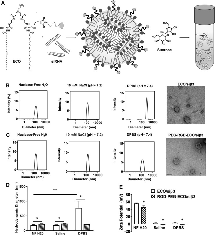
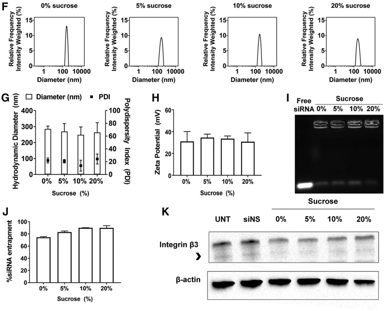
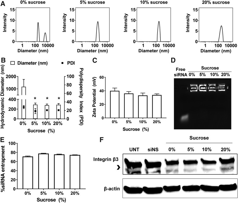
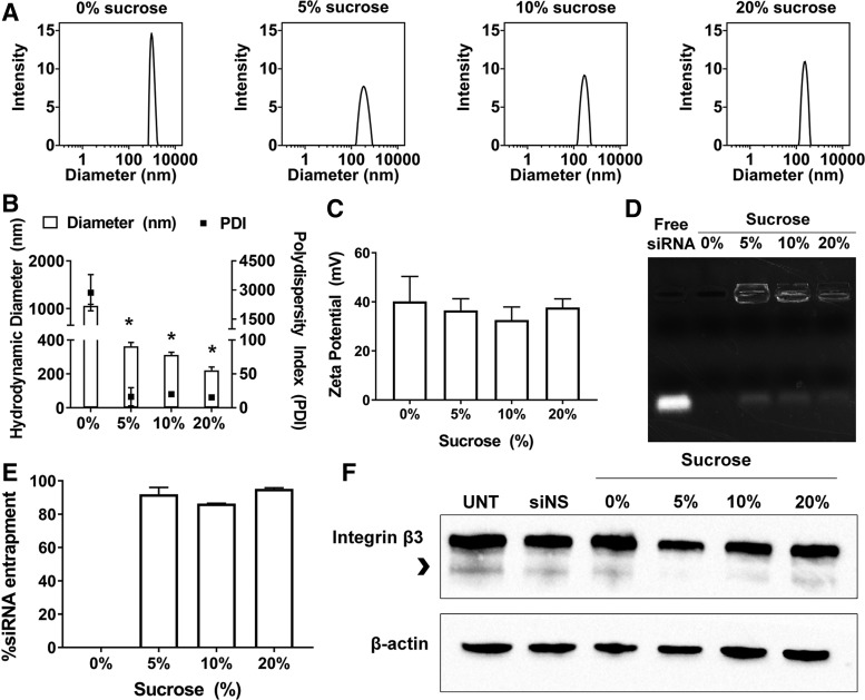
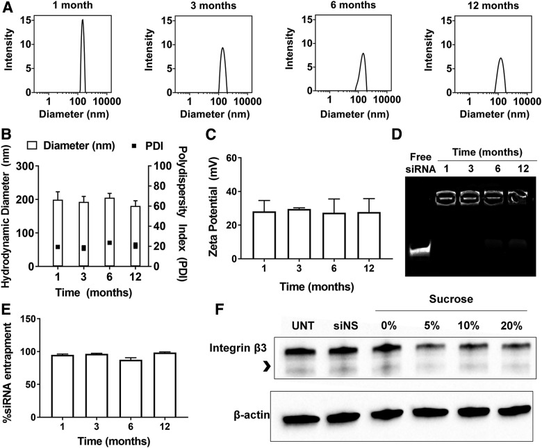
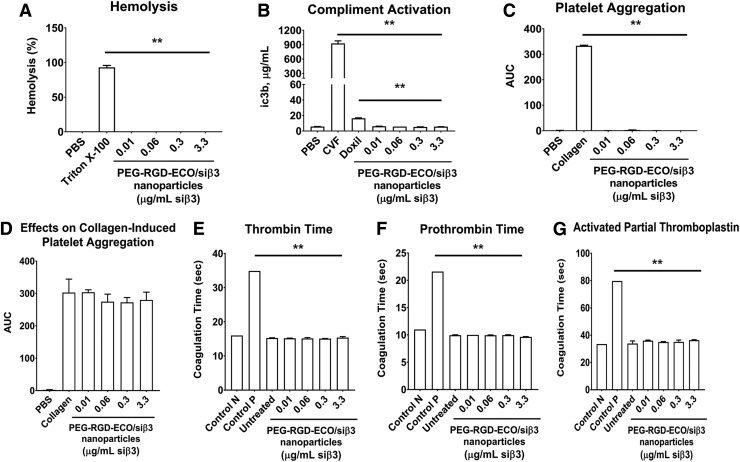
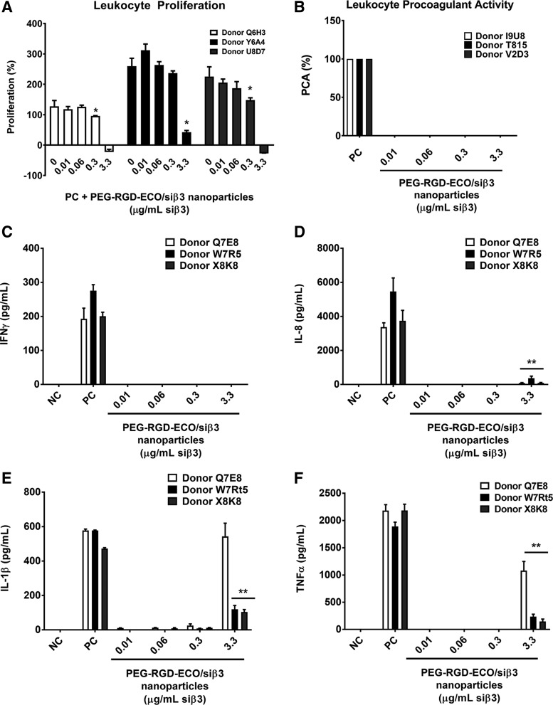
Nanoparticle based siRNA formulations often suffer from aggregation and loss of function during storage. We in this study report a frozen targeted RGD-polyethylene glycol (PEG)-ECO/siβ3 nanoparticle formulation with a prolonged shelf life and preserved nanoparticle functionality. The targeted RGD-PEG-ECO/siβ3 nanoparticles are formed by step-wised self-assembly of RGD-PEG-maleimide, ECO, and siRNA. The nanoparticles have a diameter of 224.5 ± 9.41 nm and a zeta potential to 45.96 ± 3.67 mV in water and a size of 234.34 ± 3.01 nm and a near neutral zeta potential in saline solution. The addition of sucrose does not affect their size and zeta potential and substantially preserves the integrity and biological activities of frozen and lyophilized formulations of the targeted nanoparticles. The frozen formulation with as low as 5% sucrose retains nanoparticle integrity (90% siRNA encapsulation), size distribution (polydispersity index [PDI] ≤20%), and functionality (at least 75% silencing efficiency) at -80°C for at least 1 year. The frozen RGD-PEG-ECO/siβ3 nanoparticle formulation exhibits excellent biocompatibility, with no adverse effects on hemocompatibility and minimal immunogenicity. As RNAi holds the promise in treating the previously untreatable diseases, the frozen nanoparticle formulation with the low sucrose concentration has the potential to be a delivery platform for clinical translation of RNAi therapeutics.

Keywords: ECO; RNAi; biocompatibility; long-term stability; siRNA nanoparticles.


Conflict of interest statement

Z.-R.L. is a cofounder of Cleveland Theranostics, LLC, a startup company focused on the development of multifunctional pH-sensitive amino lipids for gene therapy.

Z.-R.L. and N.R.A. have a patent interest related to this work. All other authors declare no conflicts of interest.

Figures

<b>FIG. 1.</b>
FIG. 1.
Characterization of freshly prepared ECO/siβ3 nanoparticles with and without MAL-PEG-RGD in aqueous solutions and in the presence or absence of sucrose. (A) Schematic of experimental design. Representative intensity peaks of ECO/siβ3 nanoparticles (B) and RGD-PEG-ECO/siβ3 nanoparticles (C) in nuclease-free water, 10 mM NaCl (pH = 7.2), and DPBS (CaCl2: 0.9 mM, MgCl2: 0.5 mM, KCl: 2.67 mM, KH2PO4: 1.47 mM, NaCl: 137.9 mM, Na2HPO4: 8 mM). Transmission electron microscopy images of ECO/siβ3 nanoparticles (B) and RGD-PEG-ECO/siβ3 nanoparticles (C). Comparison of the hydrodynamic diameters (D) and zeta potential (E) of ECO/siβ3 nanoparticles and RGD-PEG-ECO/siβ3 nanoparticles in aqueous solutions. (F) Representative intensity peaks of RGD-PEG-ECO/siβ3 nanoparticles containing 0%, 5%, 10%, and 20% sucrose in nuclease-free water. Hydrodynamic diameter, polydispersity index (G), and zeta potential (H) of RGD-PEG-ECO/siβ3 nanoparticles containing 0%, 5%, 10%, and 20% sucrose in nuclease-free water. (I) Agarose gel retardation of RGD-PEG-ECO/siβ3 nanoparticles containing 0%, 5%, 10%, and 20% sucrose compared to free siβ3. (J) RiboGreen assay quantifying siRNA entrapment of RGD-PEG-ECO/siβ3 nanoparticles containing 0%, 5%, 10%, and 20% sucrose. (K) Western blot analysis of β3 integrin expression (indicated by the arrowhead) in BT549 cells 48 h after RGD-PEG-ECO/siβ3 nanoparticle treatment (error bars denote SEM, *P < 0.05, **P < 0.01). DPBS, Dulbecco's phosphate-buffered saline; MAL, maleimide; PEG, polyethylene glycol; SEM, standard error of the mean.
<b>FIG. 2.</b>
FIG. 2.
Sucrose improves −80°C storage of RGD-PEG-ECO/siβ3 nanoparticles. (A) Representative intensity peaks of RGD-PEG-ECO/siβ3 nanoparticles containing 0%, 5%, 10%, and 20% sucrose poststorage in −80°C for 1 week. Comparison of the hydrodynamic diameters, polydispersity index, (B) and zeta potential (C) of RGD-PEG-ECO/siβ3 nanoparticles containing 0%, 5%, 10%, and 20% sucrose in nuclease-free water. (D) Agarose gel retardation of −80°C stored RGD-PEG-ECO/siβ3 nanoparticles containing 0%, 5%, 10%, and 20% sucrose compared with free siβ3. (E) RiboGreen assay quantifying siRNA entrapment of −80°C stored RGD-PEG-ECO/siβ3 nanoparticles containing 0%, 5%, 10%, and 20% sucrose. (F) Western blot analysis of β3 integrin (indicated by the arrowhead) expression in BT549 cells 48 h after treatment with −80°C stored RGD-PEG-ECO/siβ3 nanoparticles (error bars denote SEM, *P < 0.05 compared to formulations containing 0% sucrose).
<b>FIG. 3.</b>
FIG. 3.
Sucrose improves RGD-PEG-ECO/siβ3 nanoparticle stability postlyophilization. RGD-PEG-ECO/siβ3 nanoparticles containing 0%, 5%, 10%, and 20% sucrose were flash-frozen and lyophilized overnight. Formulations were reconstituted in their original volume of nuclease-free water. (A) Representative intensity peaks of reconstituted RGD-PEG-ECO/siβ3 nanoparticles containing 0%, 5%, 10%, and 20% sucrose. Comparison of the hydrodynamic diameters, polydispersity index, (B) and zeta potential (C) of reconstituted RGD-PEG-ECO/siβ3 nanoparticles containing 0%, 5%, 10%, and 20% sucrose in nuclease-free water. (D) Agarose gel retardation of reconstituted RGD-PEG-ECO/siβ3 nanoparticles containing 0%, 5%, 10%, and 20% sucrose compared to free siβ3. (E) RiboGreen assay quantifying siRNA entrapment of reconstituted RGD-PEG-ECO/siβ3 nanoparticles containing 0%, 5%, 10%, and 20% sucrose. (F) Western blot analysis of β3 integrin (indicated by the arrowhead) expression in BT549 cells 48 h after treatment with reconstituted RGD-PEG-ECO/siβ3 nanoparticles (error bars denote SEM, *P < 0.05 compared to formulations containing 0% sucrose).
<b>FIG. 4.</b>
FIG. 4.
Sucrose improves long-term storage of RGD-PEG-ECO/siβ3 nanoparticles. RGD-PEG-ECO/siβ3 nanoparticles containing 5% sucrose were flash-frozen and stored in −80°C for 1, 3, 6, and 12 months. (A) Representative intensity peaks of long-term stored RGD-PEG-ECO/siβ3 nanoparticles containing 5% sucrose in nuclease-free water. Comparison of the hydrodynamic diameters, polydispersity indexes (B), and zeta potential (C) of RGD-PEG-ECO/siβ3 nanoparticles stored for 1, 3, 6, and 12 months in nuclease-free water. (D) Agarose gel retardation of RGD-PEG-ECO/siβ3 nanoparticles stored for 12 months compared to free siβ3. (E) RiboGreen assay quantifying siRNA entrapment of RGD-PEG-ECO/siβ3 nanoparticles stored for 1, 3, 6, and 12 months. (F) Western blot analysis of β3 integrin expression (indicated by the arrowhead) in BT549 cells 48 h after treatment with RGD-PEG-ECO/siβ3 nanoparticles containing 0%, 5%, 10%, and 20% stored for 12 months (error bars denote SEM).
<b>FIG. 5.</b>
FIG. 5.
(A) Hemolytic activity of frozen RGD-PEG-ECO/siβ3 nanoparticles compared to negative control PBS and positive control Triton X-100. (B) Complement activation of frozen RGD-PEG-ECO/siβ3 nanoparticles compared to positive control cobra venom factor, negative control PBS, and liposomal formulation Doxil. The effect of frozen RGD-PEG-ECO/siβ3 nanoparticles on platelet aggregation (C) and collagen-induced platelet aggregation (D) was evaluated compared to positive control collagen and negative control PBS. Human plasma coagulation was evaluated comparing thrombin time (E), prothrombin time (F), and activated partial thromboplastin (G) in response to treatment with varying concentrations of frozen RGD-PEG-ECO/siβ3 nanoparticles. Controls used were normal (control N) and abnormal (control P) plasma standards, as well as untreated plasma (error bars denote SEM, **P < 0.01). PBS, phosphate-buffered saline.
<b>FIG. 6.</b>
FIG. 6.
(A) The effect of frozen RGD-PEG-ECO/siβ3 nanoparticles on PHA-M proliferation using three different donors at various concentrations compared to positive control PHA-M. (B) Leukocyte procoagulant activity of RGD-PEG-ECO/siβ3 nanoparticles compared to positive control Escherichia coli K12 lipopolysaccharide treated peripheral blood mononuclear cells. The effect of RGD-PEG-ECO/siβ3 nanoparticle treatment on pro-inflammatory cytokines IFNγ (C), IL-8 (D), IL-1β (E), and TNFα (F) was evaluated in whole blood cultures of healthy donor volunteers (n = 3) (error bars denote SEM, *P < 0.05, **P < 0.01). PHA-M, phytohemaglutinin-M; IL, interleukin; IFN, interferon; TNFα, tumor necrosis factor alpha.

References

    1. Kulkarni JA, Cullis PR. and van der Meel R. (2018). Lipid nanoparticles enabling gene therapies: from concepts to clinical utility. Nucleic Acid Ther 28:146–157 - PubMed
    1. Gaudet D, Drouin-Chartier JP. and Couture P. (2017). Lipid metabolism and emerging targets for lipid-lowering therapy. Can J Cardiol 33:872–882 - PubMed
    1. Barrett ADT. (2018). Current status of Zika vaccine development: Zika vaccines advance into clinical evaluation. NPJ Vaccines 3:24. - PMC - PubMed
    1. Adams D, Gonzalez-Duarte A, O'Riordan WD, Yang CC, Ueda M, Kristen AV, Tournev I, Schmidt HH, Coelho T, et al. (2018). Patisiran, an RNAi therapeutic, for hereditary transthyretin amyloidosis. N Engl J Med 379:11–21 - PubMed
    1. Lorenzer C, Dirin M, Winkler AM, Baumann V. and Winkler J. (2015). Going beyond the liver: progress and challenges of targeted delivery of siRNA therapeutics. J Control Release 203:1–15 - PubMed

Publication types

MeSH terms