. 2019 Aug;29(4):365-375.
doi: 10.1097/CMR.0000000000000574.

Chondroitin sulfate proteoglycan 4 enhanced melanoma motility and growth requires a cysteine in the core protein transmembrane domain

Affiliations

Chondroitin sulfate proteoglycan 4 enhanced melanoma motility and growth requires a cysteine in the core protein transmembrane domain

Jianbo Yang et al. Melanoma Res. 2019 Aug.

Abstract

Chondroitin sulfate proteoglycan 4 (CSPG4) is a cell surface proteoglycan that enhances malignant potential in melanoma and several other tumor types. CSPG4 functions as a transmembrane scaffold in melanoma cells to activate oncogenic signaling pathways such as focal adhesion kinase (FAK) and extracellular signal regulated kinases 1,2, that control motility, invasion and anchorage independent growth. Here, we demonstrate that CSPG4 promotes directional motility and anchorage independent growth of melanoma cells by organizing and positioning a signaling complex containing activated FAK to lipid rafts within the plasma membrane of migrating cells. This FAK-containing signal transduction platform, which consists of syntenin-1, active Src and caveolin-1 requires the cytoplasmic domain of CSPG4 for assembly. Enhanced directional motility promoted by this complex also requires a CSPG4 transmembrane cysteine residue C2230. Substituting C2230 with alanine (CSPG4) still permits assembly of the signaling complex, however Src remains in an inactive state. CSPG4 also fails to promote anchorage independent growth and activation of extracellular signal regulated kinases 1,2. Therapies that target the transmembrane domain of CSPG4 could be a novel strategy for limiting progression by disrupting its function as a compartmentalized motogenic and growth-promoting oncogenic signaling node.


Conflict of interest statement

CONFLICT OF INTEREST: There are no competing financial interests.

Figures

Figure 1.
Figure 1.. Truncating the CSPG4 cytoplasmic domain inhibits tumor cell migration and growth.
(A) WM1552C cells stably expressing the indicated CSPG4 variants (mock = vector control) were grown to confluence before scratching. The various colored lines represent time lapse digital tracking of an individual cell over the 24-hour period, demonstrating the relative directional motility of each cell. (B) Graphical representation of the average velocity of the individual cells tracked in A. Error bars represent SD, n= minimum of 40 cells/cell line. *p <0.01 compared to WM1552C/CSPG4WT. (C) Anchorage independent growth assay of the indicated WM1552C transfected cell lines. *p<0.001 vs. WM1552C/CSPG4WT.
Figure 2.
Figure 2.. CSPG4/β1 integrin complex formation and enhanced FAK phosphorylation.
(A) Western blot of β1 integrin from extracts of adherent cells in normal growth medium immunoprecipitated with CSPG4 antibody 9.2.27 or isotype-matched normal mouse IgG2a as indicated. (B) Western analysis of phosphorylated FAK in extracts of cells expressing full length CSPG4 or truncated variants CSPG4ΔPDZ and CSPG4ΔCD. (C) Endogenous CSPG4 and FAK co-immunoprecipitation with anti-Src antibody in parent WM1552C (CSPG4 negative) and WM1341D (CSPG4 positive) cell lines. Immunoprecipitation with normal rabbit IgG (nrIgG) was included as a negative control.
Figure 3.
Figure 3.. CSPG4 enhanced melanoma cell growth and migration requires syntenin association.
(A) Co-immunoprecipitation of syntenin with CSPG4 in the indicated cell lines. (B) CSPG4 and syntenin fail to co-precipitate in WM1552C cells expressing truncated variants CSPG4ΔPDZ or CSPG4ΔCD. (C) Co-immunoprecipitation of β1 Integrin with anti-CSPG4 antibody in WM1552C/CSPG4WT cells following knockdown of syntenin via siRNA. (D) Migration assay of fibronectin-adherent WM1552C/CSPG4WT cells following knockdown of syntenin via siRNA. Cells were transfected with syntenin siRNA 24 hours prior to plating in migration assay. *p<0.01 vs. control siRNA treatment. (E) Anchorage-independent growth assay of WM1552C/CSPG4WT cells transfected 24 hours prior to plating with syntenin siRNA. *p<0.001 vs. control siRNA treatment.
Figure 4.
Figure 4.. Transmembrane cysteine C2230 in CSPG4 is critical for enhanced cell motility and growth.
(A) Inset-Digital tracking of individual cells over the 24-hour period, demonstrating the relative directional motility of each cell. Graph -The indicated cell lines were analyzed by time-lapse digital photography as in 1A. The bars represent the average cell velocity of individually tracked cells over the 24-hour assay period, +/− S.D. (*p <0.01 compared to WM1552C/CSPG4WT) (B) Cells were serum starved for 48 hours and extracts analyzed for FAK activation (pFAKY397) by western blot. (C) Anchorage independent growth assay of the indicated WM1552C transfected cell lines. *=p<0.001 vs. WM1552C/CSPG4WT. (D) Extracts from the indicated cell lines that were either adherent (0 hr) or held in suspension for 24 hours were analyzed for total Erk1,2 and phosphorylated ERK1,2 (pErk1,2) by immunoblot.
Figure 5.
Figure 5.. Proper lipid raft localization of transmembrane cysteine C2230 in CSPG4 is essential for CSPG4-mediated Src activation.
(A) Following 24-hour induction of CSPG4 shRNA expression, 1205Lu cells that stably express a doxycycline-inducible CSPG4 shRNA were harvested (0 hr) or subsequently treated with methyl-β-cyclodextrin (10μM) for 3 hours. Extracts from these cells were isolated and analyzed by western blot to determine the expression levels of the indicated proteins. (B) and (C) Sucrose buoyant density fraction samples were collected and a portion of each fraction from the top (#3) to bottom (#8) of the density gradient from WM1552C/CSPG4C2230A (B) and WM1552C/CSPG4WT (C) were then immunoprecipitated using anti-CSPG4 antibody, electrophoresed and probed as indicated. WCL = whole cell lysate. (D) Extracts from the indicated cell lines were immunoprecipitated with anti-CSPG4 antibody. Western blots of electrophoresed samples were probed for protein expression with the indicated antibodies. Whole cell lysates from each cell line are shown for comparison.
Figure 6:
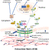
Figure 6:. CSPG4 functions as a transmembrane scaffold and oncogenic signaling node.
CSPG4 expression in tumor cells activates multiple oncogenic signaling pathways as a result of its ECM mediated scaffolding function (reviewed in [3,4,16,38]. These pathways include stimulation of Rho family GTPases to promote cytoskeletal reorganization, activation of FAK and downstream pathways to activate integrin mediated signaling, altering the transcriptome to promote mesenchymal transition and interacting with growth factor receptors to stimulate Erk 1,2 and related pathways. CSPG4 function within cholesterol rich lipid rafts requires complexing with syntenin, activated src and caveolin. Mutation of the transmembrane cysteine inhibits CSPG4 mediated FAK activation and Erk 1,2 activation. Details are described in the text.

References

    1. Fenton M, Whiteside TL, Ferrone S, Boyiadzis M. Chondroitin sulfate proteoglycan-4 (CSPG4)-specific monoclonal antibody 225.28 in detection of acute myeloid leukemia blasts. Oncol Res 2015; 22 (2):117–121. - PMC - PubMed
    1. Higgins SC, Bolteus AJ, Donovan LK, Hasegawa H, Doey L, Al Sarraj S, et al. Expression of the chondroitin sulphate proteoglycan, NG2, in paediatric brain tumors. Anticancer Res 2014; 34 (12):6919–6924. - PubMed
    1. Nicolosi PA, Dallatomasina A, Perris R. Theranostic impact of NG2/CSPG4 proteoglycan in cancer. Theranostics 2015; 5 (5):530–544. - PMC - PubMed
    1. Price MA, Colvin Wanshura LE, Yang J, Carlson J, Xiang B, Li G, et al. CSPG4, a potential therapeutic target, facilitates malignant progression of melanoma. Pigment Cell Melanoma Res 2011; 24 (6):1148–1157. - PMC - PubMed
    1. Stallcup WB. NG2 Proteoglycan Enhances Brain Tumor Progression by Promoting Beta-1 Integrin Activation in both Cis and Trans Orientations. Cancers (Basel) 2017; 9 (4). - PMC - PubMed

Publication types