. 2019 May 29;14(5):e0217236.
doi: 10.1371/journal.pone.0217236. eCollection 2019.

Lessening of porcine epidemic diarrhoea virus susceptibility in piglets after editing of the CMP-N-glycolylneuraminic acid hydroxylase gene with CRISPR/Cas9 to nullify N-glycolylneuraminic acid expression

Affiliations

Lessening of porcine epidemic diarrhoea virus susceptibility in piglets after editing of the CMP-N-glycolylneuraminic acid hydroxylase gene with CRISPR/Cas9 to nullify N-glycolylneuraminic acid expression

Ching-Fu Tu et al. PLoS One. .

Abstract

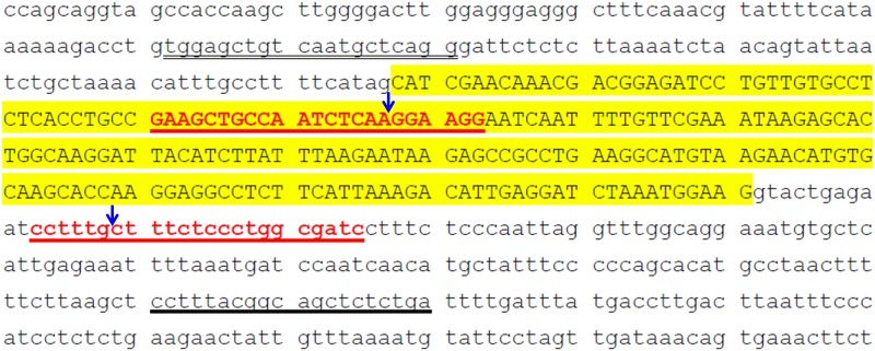
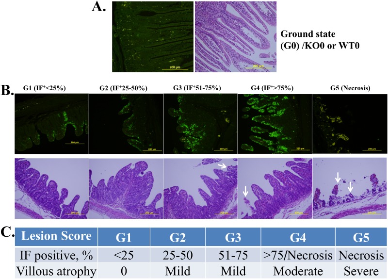
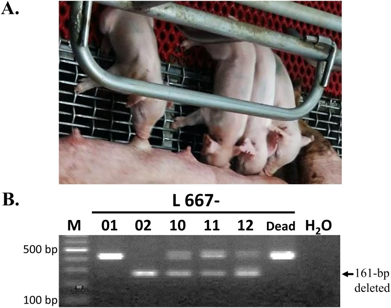
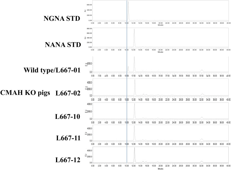
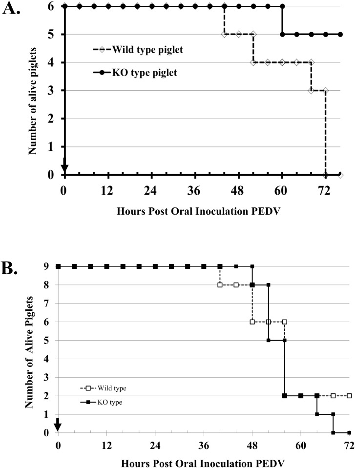
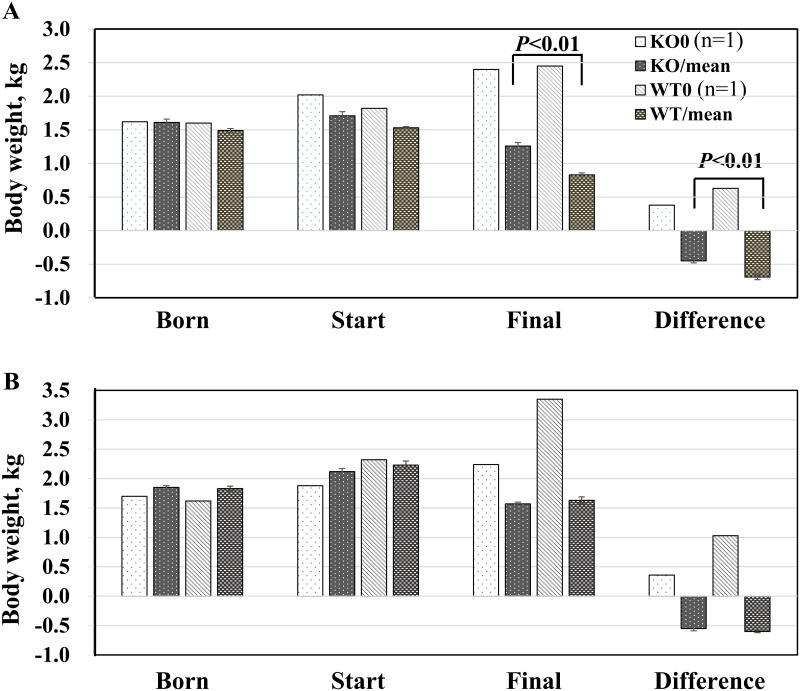
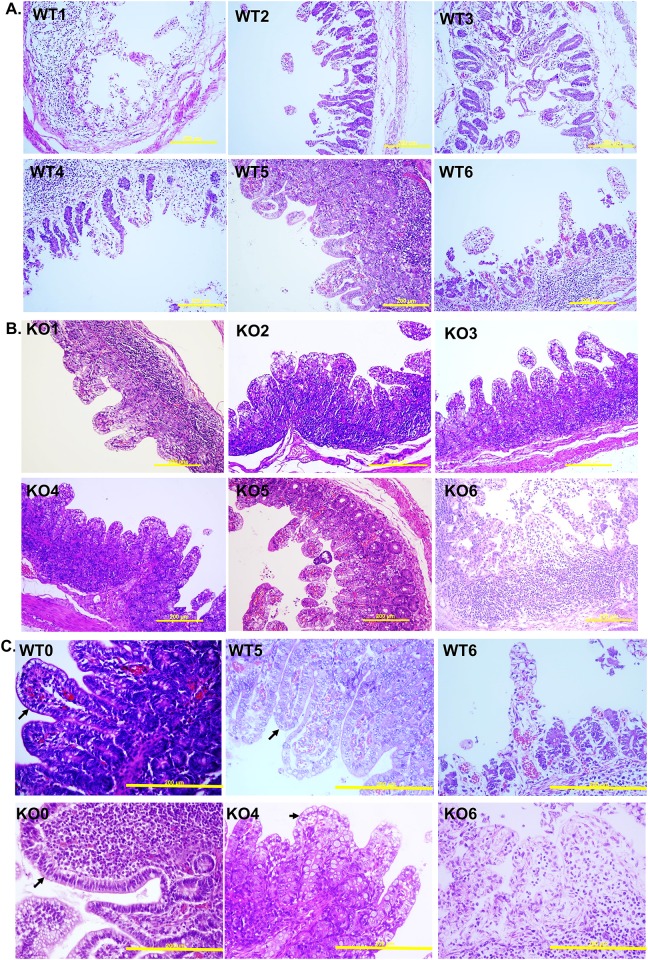
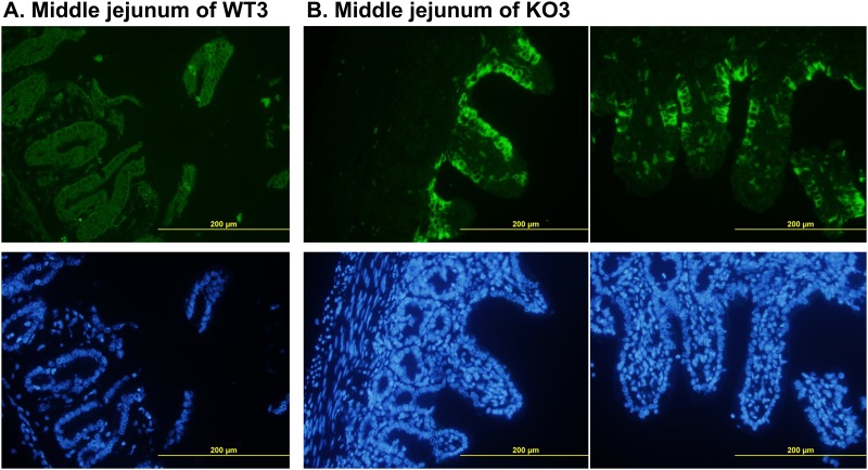
The porcine epidemic diarrhoea virus (PEDV) devastates the health of piglets but may not infect piglets whose CMP-N-glycolylneuraminic acid hydroxylase (CMAH) gene is mutated (knockouts, KO) by using CRISPR/Cas9 gene editing techniques. This hypothesis was tested by using KO piglets that were challenged with PEDV. Two single-guide RNAs targeting the CMAH gene and Cas9 mRNA were microinjected into the cytoplasm of newly fertilized eggs. Four live founders generated and proven to be biallelic KO, lacking detectable N-glycolylneuraminic acid (NGNA). The founders were bred, and homozygous offspring were obtained. Two-day-old (in exps. I, n = 6, and III, n = 15) and 3-day-old (in exp. II, n = 9) KO and wild-type (WT, same ages in respective exps.) piglets were inoculated with TCID50 1x10³ PEDV and then fed 20 mL of infant formula (in exps. I and II) or sow's colostrum (in exp. III) every 4 hours. In exp. III, the colostrum was offered 6 times and was then replaced with Ringer/5% glucose solution. At 72 hours post-PEDV inoculation (hpi), the animals either deceased or euthanized were necropsied and intestines were sampled. In all 3 experiments, the piglets showed apparent outward clinical manifestations suggesting that infection occurred despite the CMAH KO. In exp. I, all 6 WT piglets and only 1 of 6 KO piglets died at 72 hpi. Histopathology and immunofluorescence staining showed that the villus epithelial cells of WT piglets were severely exfoliated, but only moderate exfoliation and enterocyte vacuolization was observed in KO piglets. In exp. II, delayed clinical symptoms appeared, yet the immunofluorescence staining/histopathologic inspection (I/H) scores of the two groups differed little. In exp. III, the animals exhibited clinical and pathological signs after inoculation similar to those in exp. II. These results suggest that porcine CMAH KO with nullified NGNA expression are not immune to PEDV but that this KO may lessen the severity of the infection and delay its occurrence.


Conflict of interest statement

The co-author Dr. Chi-Min Chen is a virologist, especially on coronavirus and involved in the project by his responsibility to prepare the nv-PEDV and advice on the PEDV challenge procedures and the final pathological evaluation. He was our colleague but transferred to Chao Kun Biotech Ltd. This commercial affiliation did not play a role in the study design, data collection and analysis, decision to publish or preparation of the manuscript for the study. We declare that his role in this study has no conflicted interest and does not alter our adherence to PLOS ONE policies or sharing data and materials. The co-author Dr. Chien-Hong Chen was also our colleague at ATRI and was responsible for zygotes micromanipulation to generate the CMAH KO pigs. He has worked in the Reproductive Medicine Center of Lee Women’s Hospital after finished his duty in this project and without any interest conflicted to this study.

Figures

Fig 1
Fig 1. Construction of CMAH gene-edited vectors.
The porcine CMAH gene editing sites were designated on exon 2 (sense strand, red capital letters underlined in red) and intron 2 (antisense strand, red letters underlined in red). The sequences underlined in black are PCR primers (CMAH Ex2 F and CMAH Ex2 R). The sequences shown in large capital letters with yellow shading are exon 2. The blue arrows indicate the gene editing sites.
Fig 2
Fig 2. Evaluation criteria based on immunofluorescence staining and histopathological lesions (I/H score) of piglets’ intestine samples after PEDV challenge.
A. KO0 or WT0 controls for the ground state, G0. B. IF is scored as G1 to G4 based on the relative intensity of staining, whereas G5 is based on villar atrophy or defoliation observed by H/E inspection. The corresponding scores are shown in C. The arrows indicate necrotic villi; the yellow bars represent 200 μm.
Fig 3
Fig 3. Generation of CMAH gene-edited pigs.
A. Four lines of CMAH gene-edited piglets (1 male, L667-02, and 3 females, L667-10, 11, and 12) were obtained. B. CMAH KO piglets were analysed and screened by PCR. The amplicons were produced a 161-pb deleted band when two sites editing occurred simultaneously.
Fig 4
Fig 4. Genotyping by TA-cloning and sequencing of the porcine CMAH gene edited by CRISPR/Cas9 vectors directed against two sites.
A. The genotype shown displays two simultaneously mutated sites and a deleted 161-bp DNA fragment; the blue A represents an extra inserted base that appeared in L667-12. B. The indel occurred at site I of exon II of the CMAH gene. C. Details of the mutation at site II of intron 2 of the CMAH gene. The blue arrows and lines indicate the cutting site of Cas9. The blue letters represent inserted bases, and the dashed line indicates deleted bases.
Fig 5
Fig 5. Expression of NGNA/NANA in the tissues of CRISPR/Cas9 CMAH mutant founders.
L667-02, -10, -11 and -12 and their wild-type littermate (L667-01) were analysed by HPLC. NGNA STD and NANA STD are standard samples of NGNA and NANA, respectively. The blue line indicates a retention time of 10 min.
Fig 6
Fig 6. Survival of neonatal piglets after oral inoculation with nv-PEDV.
A (Exp. I), 2-day-old piglets’; and B (Exp. II), 3-day-old neonatal piglets’ survival curve after inoculated with nv-PEDV. Solid circles (A) or squares (B) with lines represent the CMAH KO piglets, and open diamonds (A) or squares (B) with dashed lines indicate wild-type piglets. The arrow shows the time of inoculation. In A at 72 hpi, three moribund WT piglets are classified as dead piglets.
Fig 7
Fig 7. Body weights of neonatal piglets before and after oral PEDV inoculation.
A. 2-day-old piglets (n = 6) and B. 3-day-old piglets (n = 9) in Exp. I and Exp. II, respectively, show body weights changes 72 h after inoculated with nv-PEDV. K0 and W0 represent KO and WT animals that were not inoculated with PEDV and were reared by their dams on the farm. KO and WT are knockout treated and wild-type treated animals, respectively.
Fig 8
Fig 8. Pathological inspection of piglets’ intestine at the middle jejunum by H/E staining.
Panels A. and B. indicate wild-type and knockout piglets, respectively, after PEDV oral inoculation. C. The samples from control, the best and the worst pathological responded piglets, which are no nv-PEDV-inoculated, survival and dead, respectively, at 72 hpi. The upper panels (WT0, WT5 and WT6) are samples from wild type piglets and the down panels (KO0, KO4 and KO6 are samples from CMAH KO piglets. Arrows indicate epithelial cells and the yellow bars indicate 200 μm.
Fig 9
Fig 9. Immunofluorescence staining with an antibody against nv-PEDV N protein.
WT3 (A) shows a sample from a wild-type piglet, and KO3 (B) shows a sample from a double-chromosome CMAH gene knockout piglet. The samples shown in the upper, green colour, and lower, blue colour, panels are fluorescent stained with nv-PEDV antibody and DAPI, respectively. The yellow bars indicate 200 μm.

References

    1. Lee C. Porcine epidemic diarrhea virus: an emerging and re-emerging epizootic swine virus. Virol J. 2015;12: 193 10.1186/s12985-015-0421-2 - DOI - PMC - PubMed
    1. Pensaert MB, de Bouck P. A new coronavirus-like particle associated with diarrhea in swine. Arch Virol. 1978;58: 243–247. - PMC - PubMed
    1. Chae C, Kim O, Choi C, Min K, Cho WS, Kim J, et al. Prevalence of Porcine epidemic diarrhoea virus and transmissible gastroenteritis virus infection in Korean pigs. Vet Rec. 2000;147: 606–608. - PubMed
    1. Sun RQ, Cai RJ, Chen YQ, Liang PS, Chen DK, Song CX. Outbreak of Porcine epidemic diarrhea in suckling piglets, China. Emerg Infect Dis. 2012;18: 161–163. 10.3201/eid1801.111259 - DOI - PMC - PubMed
    1. Lee S, Lee C. Outbreak-related Porcine epidemic diarrhea virus strains similar to US strains, South Korea, 2013. Emerg Infect Dis. 2014;20: 1223–1226. 10.3201/eid2007.140294 - DOI - PMC - PubMed

Publication types

MeSH terms

Substances