. 2019 May 28;20(11):2612.
doi: 10.3390/ijms20112612.

Deoxypodophyllotoxin Exerts Anti-Cancer Effects on Colorectal Cancer Cells Through Induction of Apoptosis and Suppression of Tumorigenesis

Affiliations

Deoxypodophyllotoxin Exerts Anti-Cancer Effects on Colorectal Cancer Cells Through Induction of Apoptosis and Suppression of Tumorigenesis

Chathurika D B Gamage et al. Int J Mol Sci. .

Abstract

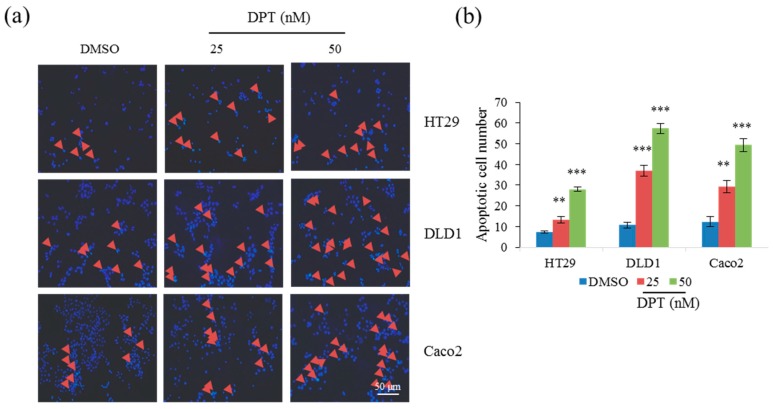
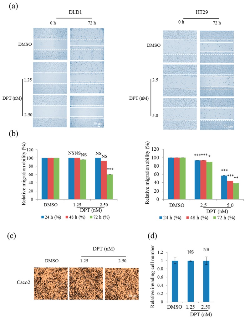
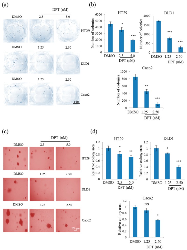
Deoxypodophyllotoxin (DPT) is a cyclolignan compound that exerts anti-cancer effects against various types of cancers. DPT induces apoptosis and inhibits the growth of breast, brain, prostate, gastric, lung, and cervical tumors. In this study, we sought to determine the effect of DPT on cell proliferation, apoptosis, motility, and tumorigenesis of three colorectal cancer (CRC) cell lines: HT29, DLD1, and Caco2. DPT inhibited the proliferation of these cells. Specifically, the compound-induced mitotic arrest in CRC cells by destabilizing microtubules and activating the mitochondrial apoptotic pathway via regulation of B-cell lymphoma 2 (Bcl-2) family proteins (increasing Bcl-2 associated X (BAX) and decreasing B-cell lymphoma-extra-large (Bcl-xL)) ultimately led to caspase-mediated apoptosis. In addition, DPT inhibited tumorigenesis in vitro, and in vivo skin xenograft experiments revealed that DPT significantly decreased tumor size and tumor weight. Taken together, our results suggest DPT to be a potent compound that is suitable for further exploration as a novel chemotherapeutic for human CRC.

Keywords: apoptosis; colorectal cancer; deoxypodophyllotoxin; mitotic arrest; tubulin polymerization; tumorigenic potentials.


Conflict of interest statement

The authors declare no conflict of interest.

Figures

Figure 1
Figure 1
Cytotoxic effects of podophyllotoxin, picropodophyllotoxin, and deoxypodophyllotoxin (DPT) in CRC cells. Cells were treated for 48 h with podophyllotoxin (a) and picropodophyllotoxin (b) at concentrations from 100 to 300 nM, and DPT (c) at concentrations from 10 to 50 nM. Cell viability was measured using an MTT assay. Data represent means ± S.E.M.; n = 3. * p < 0.05; ** p < 0.01; *** p < 0.001; NS, no significant difference compared with the DMSO-treated group.
Figure 2
Figure 2
Induction of apoptosis in CRC cells by DPT, as determined by staining with either Hoechst (a,b) or propidium iodide (PI)/Annexin V–FITC (c,d). (a) Hoechst staining of CRC cells treated with 25 or 50 nM DPT for 12 h. Arrowheads indicate nuclear condensation in cells. (b) Quantification of cells with condensed nuclei in Hoechst stained cells treated with 25 or 50 nM DPT. (c) PI and Annexin V–FITC staining of CRC cells treated with 25 or 50 nM DPT for 48 h. (d) Quantification of apoptosis in PI/Annexin V-FITC stained cells treated with 25 or 50 nM DPT. Results are representative of three experiments. Data represent mean ± S.E.M. ** p < 0.01; *** p < 0.001 compared with the DMSO-treated group.
Figure 2
Figure 2
Induction of apoptosis in CRC cells by DPT, as determined by staining with either Hoechst (a,b) or propidium iodide (PI)/Annexin V–FITC (c,d). (a) Hoechst staining of CRC cells treated with 25 or 50 nM DPT for 12 h. Arrowheads indicate nuclear condensation in cells. (b) Quantification of cells with condensed nuclei in Hoechst stained cells treated with 25 or 50 nM DPT. (c) PI and Annexin V–FITC staining of CRC cells treated with 25 or 50 nM DPT for 48 h. (d) Quantification of apoptosis in PI/Annexin V-FITC stained cells treated with 25 or 50 nM DPT. Results are representative of three experiments. Data represent mean ± S.E.M. ** p < 0.01; *** p < 0.001 compared with the DMSO-treated group.
Figure 3
Figure 3
Induction of mitotic arrest in human CRC cells and inhibition of tubulin polymerization by DPT. (a) Flow cytometric analysis of the cell-cycle distribution of Caco2 (48 h) and DLD1 (24 h) cells after treatment with DPT at concentrations ranging from nontoxic to toxic (1.25–25 nM). (b) Effects of DPT at 50 nM, 5 µM, and 10 µM on the in vitro polymerization of purified tubulin. The effect of DPT was examined in a GTP-containing buffer. Results are representative of three experiments.
Figure 4
Figure 4
Activation of apoptosis by DPT in CRC cells. (a,b,d) Western blot of poly(ADP-ribose) polymerase (PARP) and caspase-3 (a), BAX (b), and Bcl-xL (d) in cells treated with 25 or 50 nM DPT. (c,e) Quantification of Bax (c) and Bcl-XL (e) protein levels in cells treated with DPT. Data represent means ± S.E.M. * p < 0.05; ** p < 0.01; *** p < 0.001; NS, no significant difference compared with the DMSO-treated group.
Figure 5
Figure 5
Inhibition of CRC cell motility by DPT. (a,b) Migration assay of DLD1 and HT29 cells treated with 1.25, 2.5, or 5 nM DPT (a), and quantitative analysis of wound length (b). (c,d) Invasion assays in Caco2 cells treated with 1.25 or 2.5 nM DPT; (d) shows quantitation of invading cell numbers in each group. Representative images from three independent experiments are shown in (c). Data represent means ± S.E.M. * p < 0.05; ** p < 0.01; *** p < 0.001; NS, no significant difference relative to DMSO-treated Caco2 cells.
Figure 6
Figure 6
Inhibition of proliferation and anchorage-independent growth of HT29, DLD1, and Caco2 cells using sub-lethal concentrations of DPT. (a,b) Clonogenic assay of HT29, DLD1, and Caco2 cells treated with DPT (a) and quantification of colony number in each group (b). (c,d) Soft-agar colony-formation assays of HT29, DLD1, and Caco2 cells treated with DPT (c), and quantification of percent colony area in each group (d). Representative images are shown from three independent experiments. Data represent means ± S.E.M.; * p < 0.05; ** p < 0.01; *** p < 0.001; NS, no significant difference compared with the DMSO-treated group.
Figure 7
Figure 7
Inhibition of in vivo tumorigenesis by DPT in a xenograft mouse model. (a) Images of tumor tissues, representative of seven mice from the DPT-treated group and DMSO-treated control group. (b,c) Quantitative analysis of isolated tumor size (b) and tumor weight (c) for each group. (d) Body weight of the control and DPT-treated mice. Results are reported as means ± S.E.M.; ** p < 0.01; NS, no significant difference compared with the DMSO-treated group.

References

    1. Arnold M., Sierra M.S., Laversanne M., Soerjomataram I., Jemal A., Bray F. Global patterns and trends in colorectal cancer incidence and mortality. Gut. 2017;66:683–691. doi: 10.1136/gutjnl-2015-310912. - DOI - PubMed
    1. PDQ Adult Treatment Editorial Board Colon cancer treatment (PDQ(R)): Health professional version. [(accessed on 22 February 2019)];PDQ Cancer Information Summaries. Available online: https://www.ncbi.nlm.nih.gov/books/NBK65858/
    1. Tian Z.H., Chu Y.Y., Wang H., Zhong L.L., Deng M.Y., Li W.B. Biological activity and interaction mechanism of the diketopiperazine derivatives as tubulin polymerization inhibitors. RSC Adv. 2018;8:1055–1064. doi: 10.1039/C7RA12173C. - DOI - PMC - PubMed
    1. Xi J., Zhu X., Feng Y., Huang N., Luo G., Mao Y., Han X., Tian W., Wang G., Luo R., et al. Development of a novel class of tubulin inhibitors with promising anticancer activities. Mol. Cancer Res. 2013;11:856–864. doi: 10.1158/1541-7786.MCR-12-0177. - DOI - PubMed
    1. Sakchaisri K., Kim S.O., Hwang J., Soung N.K., Lee K.H., Choi T.W., Lee Y., Park C.M., Thimmegowda N.R., Lee P.Y., et al. Anticancer activity of a novel small molecule tubulin inhibitor stk899704. PLoS ONE. 2017;12:e0173311. doi: 10.1371/journal.pone.0173311. - DOI - PMC - PubMed

MeSH terms