. 2019 May 29;20(1):293.
doi: 10.1186/s12859-019-2902-6.

Pairwise visual comparison of small RNA secondary structures with base pair probabilities

Affiliations

Pairwise visual comparison of small RNA secondary structures with base pair probabilities

Serge Léger et al. BMC Bioinformatics. .

Abstract

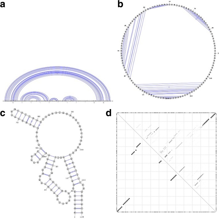
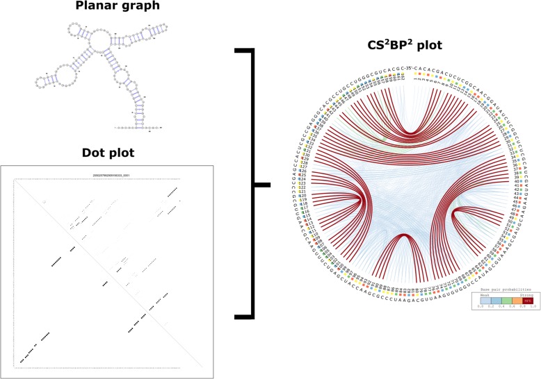
Background: Predicted RNA secondary structures are typically visualized using dot-plots for base pair binding probabilities and planar graphs for unique structures, such as the minimum free energy structure. These are however difficult to analyze simultaneously.

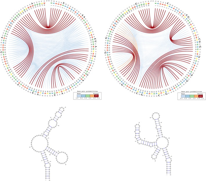
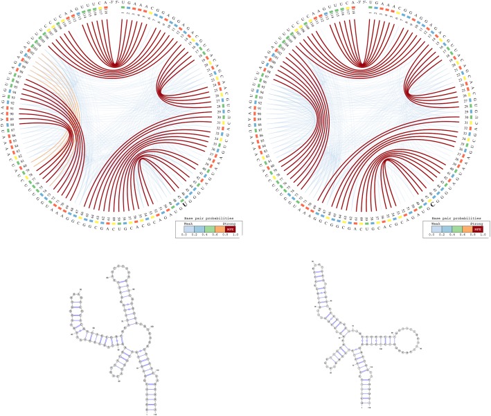
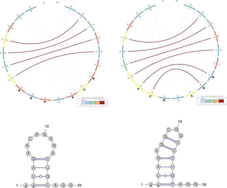
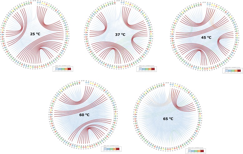
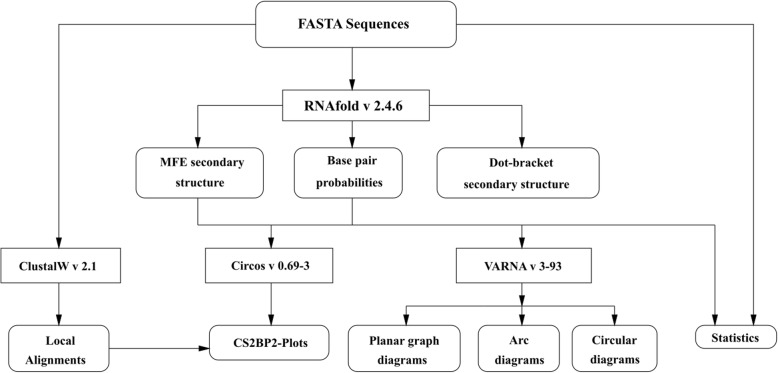
Results: This work introduces a compact unified view of the most stable conformation of an RNA secondary structure and its base pair probabilities, which is called the Circular Secondary Structure Base Pairs Probabilities Plot (CS2BP2-Plot). Along with our design we provide access to a web server implementation of our solution that facilitates pairwise comparison of short RNA (and DNA) sequences up to 200 base pairs. The web server first calculates the minimum free energy secondary structure and the base pair probabilities for up to 10 RNA or DNA sequences using RNAfold and then provides a two panel comparative view that includes CS2BP2-Plots along with the traditional graph, planar and circular diagrams obtained with VARNA. The CS2BP2-Plots include highlighting of the nucleotide differences between two selected sequences using ClustalW local alignments. We also provide descriptive statistics, dot-bracket secondary structure representations and ClustalW local alignments for compared sequences.

Conclusions: Using circular diagrams and colour and weight-coded arcs, we demonstrate how a single image can replace the state-of-the-art dual representations (dot-plots and minimum free energy structures) for base-pair probabilities of RNA secondary structures while allowing efficient exploration and comparison of different RNA conformations via a web server front end. With that, we provide the community, especially the biologically oriented, with an intuitive tool for ncRNA visualization. Web-server: https://nrcmonsrv01.nrc.ca/cs2bp2plot.


Conflict of interest statement

The authors declare that they have no competing interests.

Figures

Fig. 1
Fig. 1
Different representations of an RNA secondary structure. Sub-figure (a) depicts a linear arc diagram, sub-figure (b) a circular diagram, sub-figure (c) a planar graph, and sub-figure (d) a dot-plot diagram for the ancestral ncRNA region of HAR1
Fig. 2
Fig. 2
CS2BP2-Plot (top) and planar graphs (bottom) for ancestral (left) and Denisovan (right) HAR1 secondary structures. The CS2BP2-Plots include sequence differences highlighted in bold. The sequence differences are highlighted after we infer them from a ClustalW pairwise local alignment
Fig. 3
Fig. 3
- CS2BP2-Plot (top) and planar graphs (bottom) for Denisovan (left) and Human (right) HAR1 secondary structures. The CS2BP2-Plots include sequence differences highlighted in bold
Fig. 4
Fig. 4
The RNAbows (top) and CS2BP2-Plots (bottom) representations for the human and ancestral HAR1 secondary structures. The RNAbows arc diagram was obtained using the diffRNABow functionality available at http://rna.williams.edu/rnabows/ using the Vienna-2.0.2 folding method. The CS2BP2-Plots were obtained using the default parameter settings available at https://nrcmonsrv01.nrc.ca/cs2bp2plot/
Fig. 5
Fig. 5
The CS2BP2-Plots (top) and planar graphs (bottom) for secondary structures of a small bi-stable RNA. Sub-figures (a) and (d) describe the secondary structure conformations at 25 °C. Sub-figures (b) and (e) depict the secondary structure conformations at 70 °C and sub-figures (c) and (f) depict the secondary structure conformations at 75 °C
Fig. 6
Fig. 6
Active (left) and inactive (right) gRNAs from Thyme et al., 2016. The secondary structures of the gRNAs include a pair of non-canonical base pairs (G-U) at positions 3–16
Fig. 7
Fig. 7
The CS2BP2-Plots and planar graph diagrams of the sequence flanking an SNV in the 3’UTR of SUB1. The activated RNA polymerase II transcriptional co-activator p15 or SUB1 homolog was previously identified as a riboSNitch by Corley et al. [24]
Fig. 8
Fig. 8
CS2BP2-Plots for ailA putative virulent factor in Yersinia pseudotuberculosis at five different temperatures. The ailA putative virulent factor from Y. pseudotuberculosis shows significantly different layouts of their secondary structures at 5 different temperatures: 25 °C, 37 °C, 45 °C, 60 °C and 65 °C
Fig. 9
Fig. 9
The planar graph, dot plot and CS2BP2-Plot of the nucleotide sequence of wheat-embryo cytosol 5.8S ribosomal RNA. The CS2BP2-Plot includes information typically included in two separate graphical representations (a planar graph for the minimum free energy base pairing configuration and a dot-plot for base pair probabilities)
Fig. 10
Fig. 10
CS2BP2-Plot web server functionality flowchart. The web-server pipeline that implements and automatically generates the CS2BP2-Plots is depicted here. All data files (input and output) are depicted as round boxes, while the software used to process the files are described with rectangles

References

    1. Mortimer SA, Kidwell MA, Doudna JA. Insights into RNA structure and function from genome-wide studies. Nat Rev Genet. 2014;15:469–479. doi: 10.1038/nrg3681. - DOI - PubMed
    1. Cruz JA, Westhof E. The dynamic landscapes of RNA architecture. Cell. 2009;136:604–609. doi: 10.1016/j.cell.2009.02.003. - DOI - PubMed
    1. Darty K, Denise A, Ponty Y. VARNA: interactive drawing and editing of the RNA secondary structure. Bioinformatics. 2009;25:1974–1975. doi: 10.1093/bioinformatics/btp250. - DOI - PMC - PubMed
    1. Aalberts DP, Jannen WK. Visualizing RNA base-pairing probabilities with RNAbow diagrams. RNA. 2013;19:475–478. doi: 10.1261/rna.033365.112. - DOI - PMC - PubMed
    1. Lai D, Proctor JR, Zhu JYA, Meyer IM. R-CHIE: a web server and R package for visualizing RNA secondary structures. Nucleic Acids Res. 2012;40:e95. doi: 10.1093/nar/gks241. - DOI - PMC - PubMed