. 2019 May 29;7(1):85.
doi: 10.1186/s40478-019-0734-2.

Gerstmann-Sträussler-Scheinker disease revisited: accumulation of covalently-linked multimers of internal prion protein fragments

Affiliations

Gerstmann-Sträussler-Scheinker disease revisited: accumulation of covalently-linked multimers of internal prion protein fragments

Laura Cracco et al. Acta Neuropathol Commun. .

Erratum in

Abstract

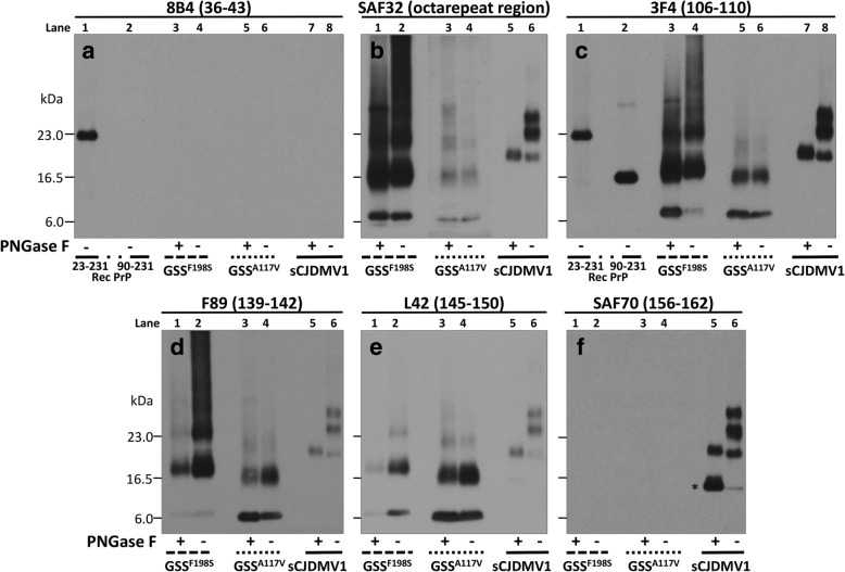
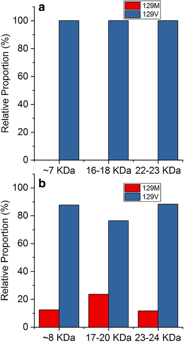
Despite their phenotypic heterogeneity, most human prion diseases belong to two broadly defined groups: Creutzfeldt-Jakob disease (CJD) and Gerstmann-Sträussler-Scheinker disease (GSS). While the structural characteristics of the disease-related proteinase K-resistant prion protein (resPrPD) associated with the CJD group are fairly well established, many features of GSS-associated resPrPD are unclear. Electrophoretic profiles of resPrPD associated with GSS variants typically show 6-8 kDa bands corresponding to the internal PrP fragments as well as a variable number of higher molecular weight bands, the molecular nature of which has not been investigated. Here we have performed systematic studies of purified resPrPD species extracted from GSS cases with the A117V (GSSA117V) and F198S (GSSF198S) PrP gene mutations. The combined analysis based on epitope mapping, deglycosylation treatment and direct amino acid sequencing by mass spectrometry provided a conclusive evidence that high molecular weight resPrPD species seen in electrophoretic profiles represent covalently-linked multimers of the internal ~ 7 and ~ 8 kDa fragments. This finding reveals a mechanism of resPrPD aggregate formation that has not been previously established in prion diseases.

Keywords: Aggregate formation; Creutzfeldt-Jakob disease; Epitope mapping; Mass spectrometry; Multimers; Prion protein.


Conflict of interest statement

The authors declare that they have no competing interests.

Figures

Fig. 1
Fig. 1
Epitope mapping combined with deglycosylation of purified resPrPD associated with GSSF198S, GSSA117V and sCJDMV1 used as control. PNGase F-treated or untreated, PK-resistant PrPD (resPrPD) purified (see Materials and Methods) from GSSF198S, GSSA117V and sCJDMV1 control were blotted and probed with the Abs to PrP denoted, with their epitopes, at the top of each panel. Panel a: Only full-length (PK-untreated) recombinant PrP (23–231) was detected by this Ab to the proximal N-terminal region confirming the absence of resPrPD with complete N-terminus. Panels b to e: Both GSSF198S and GSSA117V resPrPD conformers were selectively detected by the same Abs to PrP N- and C-terminal regions; both also showed no major variation of the resPrPD banding pattern following deglycosylation indicating that most of the resPrPD is unglycosylated in both conditions. Although overall similar, the two resPrPD profiles differed especially in the higher molecular weight region suggesting a distinct repertoire, or relative proportions, of polymers in the two GSS variants. The GSS banding patterns clearly differed from the three-band pattern of sCJDMV1, two of which are glycosylated (See Results for detailed description). Panel c includes the bands of both 23–231 and 90–231 recombinant PrP which have been used as molecular weight markers. In b the portion of the panel with the GSSA117V samples required longer exposure. In panel f, * indicates the 13 kDa component of the glycosylated and anchor bearing PrP 12/13 C-terminus fragments with N-termini at residues 162–167 and 154–156, respectively [24, 41]
Fig. 2
Fig. 2
Mass spectrometry (MS)-based sequencing of PrP fragments in the material extracted from individual gel bands of two GSS variants. GSSA117V resPrPD (a) and GSSF198S resPrPD (b). The material present in these bands was subjected to trypsin digestion, followed by MS analysis of tryptic fragments. The fragments identified by MS for species extracted from individual gel bands are shown as blue, red and green lines. Amino acid sequence within the 70-152 region is shown above the lines. Residues marked in red represent potential cleavage sites in the relevant region of PrP; 129M/V polymorphic residues are marked in blue
Fig. 3
Fig. 3
Relative abundance of 129M and 129V PrP variants in resPrPD associated with GSSA117V and GSSF198S. a: GSSA117V; b: GSSF198S. The relative abundance of resPrPD with M or V at residue 129 reflects the representation of the PrP mutation, which is coupled with the 129V in both GSS variants. Approximately 10–25% of resPrPD could be identified as non-mutated (wild type) in GSSF198S while only mutated resPrPD could be detected in GSSA117V. The relative populations were determined by mass spectrometry using the spectral counting method

References

    1. Aguilar-Calvo P, Xiao X, Bett C, Erana H, Soldau K, Castilla J, Nilsson KP, Surewicz WK, Sigurdson CJ. Post-translational modifications in PrP expand the conformational diversity of prions in vivo. Sci Rep. 2017;7:43295. doi: 10.1038/srep43295. - DOI - PMC - PubMed
    1. Al-Hilaly YK, Biasetti L, Blakeman BJ, Pollack SJ, Zibaee S, Abdul-Sada A, Thorpe JR, Xue WF, Serpell LC. The involvement of dityrosine crosslinking in alpha-synuclein assembly and deposition in Lewy bodies in Parkinson's disease. Sci Rep. 2016;6:39171. doi: 10.1038/srep39171. - DOI - PMC - PubMed
    1. Al-Hilaly YK, Williams TL, Stewart-Parker M, Ford L, Skaria E, Cole M, Bucher WG, Morris KL, Sada AA, Thorpe JR, Serpell LC. A central role for dityrosine crosslinking of amyloid-beta in Alzheimer's disease. Acta Neuropathol Commun. 2013;1:83. doi: 10.1186/2051-5960-1-83. - DOI - PMC - PubMed
    1. Bantscheff M, Lemeer S, Savitski MM, Kuster B. Quantitative mass spectrometry in proteomics: critical review update from 2007 to the present. Anal Bioanal Chem. 2012;404:939–965. doi: 10.1007/s00216-012-6203-4. - DOI - PubMed
    1. Baskakov IV, Caughey B, Requena JR, Sevillano AM, Surewicz WK, Wille H. The prion 2018 round tables (I): the structure of PrP(Sc) Prion. 2019;13:46–52. doi: 10.1080/19336896.2019.1569450. - DOI - PMC - PubMed

Publication types

MeSH terms