Review
. 2019 May 29;32(3):e00138-18.
doi: 10.1128/CMR.00138-18. Print 2019 Jun 19.

Cystic Fibrosis and Pseudomonas aeruginosa: the Host-Microbe Interface

Affiliations
Review

Cystic Fibrosis and Pseudomonas aeruginosa: the Host-Microbe Interface

Sankalp Malhotra et al. Clin Microbiol Rev. .

Abstract

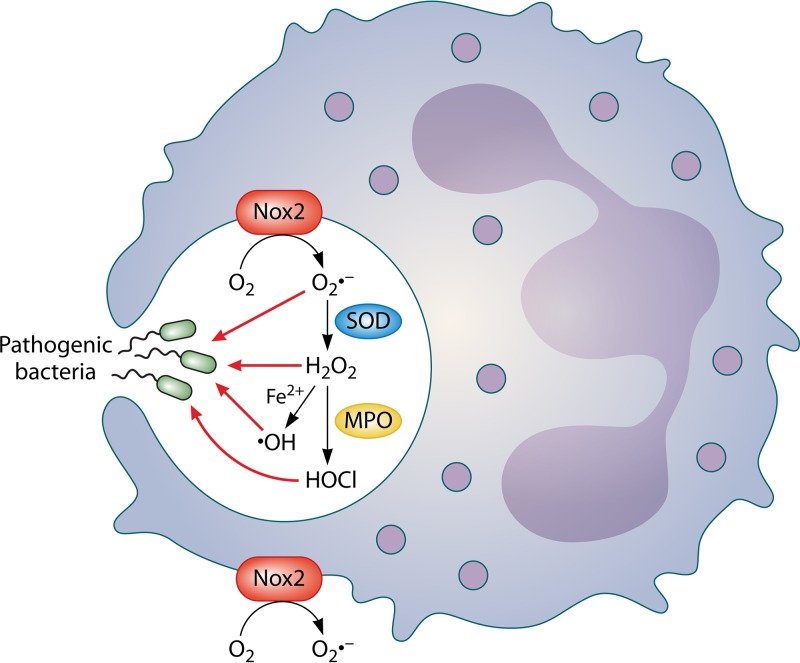
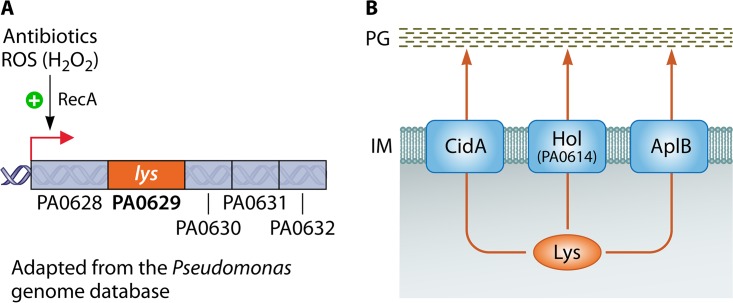
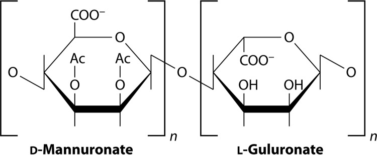
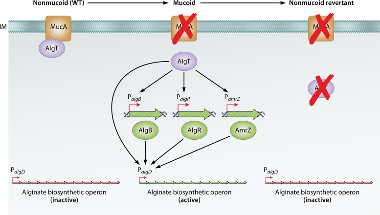
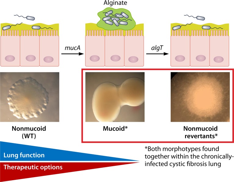
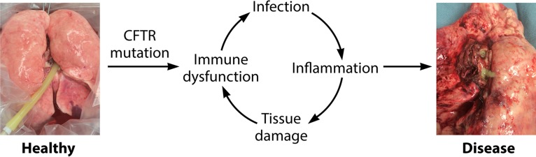
In human pathophysiology, the clash between microbial infection and host immunity contributes to multiple diseases. Cystic fibrosis (CF) is a classical example of this phenomenon, wherein a dysfunctional, hyperinflammatory immune response combined with chronic pulmonary infections wreak havoc upon the airway, leading to a disease course of substantial morbidity and shortened life span. Pseudomonas aeruginosa is an opportunistic pathogen that commonly infects the CF lung, promoting an accelerated decline of pulmonary function. Importantly, P. aeruginosa exhibits significant resistance to innate immune effectors and to antibiotics, in part, by expressing specific virulence factors (e.g., antioxidants and exopolysaccharides) and by acquiring adaptive mutations during chronic infection. In an effort to review our current understanding of the host-pathogen interface driving CF pulmonary disease, we discuss (i) the progression of disease within the primitive CF lung, specifically focusing on the role of host versus bacterial factors; (ii) critical, neutrophil-derived innate immune effectors that are implicated in CF pulmonary disease, including reactive oxygen species (ROS) and antimicrobial peptides (e.g., LL-37); (iii) P. aeruginosa virulence factors and adaptive mutations that enable evasion of the host response; and (iv) ongoing work examining the distribution and colocalization of host and bacterial factors within distinct anatomical niches of the CF lung.

Keywords: ROS; airway; antimicrobial peptides; cystic fibrosis; inflammation; innate immunity; lung infection; reactive oxygen species.


Figures

FIG 1
FIG 1
Simplified model for intracellular and extracellular generation of reactive oxygen species (ROS) by phagocytes. Molecular oxygen is converted by NADPH oxidase 2 (Nox2) to superoxide (O2·−). O2·− is converted to hydrogen peroxide (H2O2) by superoxide dismutase (SOD). H2O2 may then be converted to either hydroxyl free radical (·OH) (via Fenton chemistry in the presence of ferrous iron [Fe2+]) or halogenated ROS, such as hypochlorous acid (HOCl), via myeloperoxidase (MPO). Nox2 is localized within phagosomes as well as on the cell membrane (158).
FIG 2
FIG 2
Working model for Lys-mediated autolysis in P. aeruginosa. (A) Part of the R/F-pyocin gene cluster (PA0628-PA0632), a putative operon containing lys (PA0629), which encodes an endolysin implicated in P. aeruginosa autolysis (“explosive cell lysis”) and eDNA release. As part of the RecA-dependent SOS response, lys transcription is upregulated by cell membrane and genotoxic stresses (caused by exposure to antibiotics or reactive oxygen species [e.g., H2O2]) (103, 336). (B) Lys traverses the inner membrane (IM) via holin proteins (CidB, AplB, and a putative holin, Hol [PA0614]) and degrades peptidoglycan (PG), thereby destabilizing the cell wall and prompting cell lysis (103).
FIG 3
FIG 3
Predominant colony morphologies of P. aeruginosa variants within the cystic fibrosis lung. P. aeruginosa strains were grown and imaged on Vogel-Bonner minimal medium (VBMM) with Congo Red. (A) Wild type (nonmucoid); (B) rugose small-colony variant (RSCV) (nonmucoid); (C) mucoid variant. RSCVs overproduce Psl and Pel exopolysaccharides. Mucoid variants overproduce the alginate exopolysaccharide. The red color of RSCV colonies is due to positive staining with Congo Red. All colonies were imaged at the same magnification. (Adapted from reference with permission.)
FIG 4
FIG 4
Chemical structure of alginate. Alginate is a polyanionic exopolysaccharide, composed of β-1,4-linked d-mannuronic and l-guluronic acids. (Reproduced from reference with permission.)
FIG 5
FIG 5
Regulation of alginate biosynthesis: mucoid conversion and reversion. In wild-type, nonmucoid P. aeruginosa, the alginate biosynthetic operon (algD-algA) is inactive, as a sigma factor, AlgT, is sequestered at the inner membrane (IM) by its cognate anti-sigma factor, MucA. Acquisition of a mucA mutation results in a truncated MucA protein that is no longer able to bind AlgT. AlgT can then activate alginate biosynthesis in 2 principal ways: (i) by activating the transcription of the alginate biosynthetic operon at the algD promoter (PalgD) and (ii) by activating the transcription of genes encoding three ancillary transcription factors, AlgB, AlgR, and AmrZ, which also bind to PalgD and are essential for alginate production. Mucoid variants of P. aeruginosa can revert back to a nonmucoid phenotype via the acquisition of a secondary (suppressor) mutation in algT, which inactivates alginate biosynthesis (375, 425).
FIG 6
FIG 6
Paradigm for mucoid conversion and reversion within the cystic fibrosis (CF) lung. CF patients are initially infected by environmental (wild-type [WT]) isolates of P. aeruginosa. During chronic infection of the CF lung, nonmucoid variants of P. aeruginosa commonly acquire a mucA mutation, leading to a phenotypic switch to mucoidy (i.e., alginate overproduction). Mucoid conversion portends a decline in patient lung function, and mucoid variants exhibit increased recalcitrance to antibiotic therapy. However, via the acquisition of second-site, suppressor mutations (e.g., algT), mucoid variants commonly revert back to a nonmucoid phenotype both in vitro and in vivo. Mixed populations of mucoid and nonmucoid revertants of P. aeruginosa are commonly observed within the CF lung, suggesting a selective advantage for mucoid/nonmucoid coinfection. (Adapted from reference with permission.)
FIG 7
FIG 7
The pathogenesis of cystic fibrosis pulmonary disease: a vicious cycle driven by immune dysfunction and bacterial infection. Although the newborn CF lung is histopathologically comparable to a healthy lung, the CFTR mutation causes significant, primary defects in innate immunity (e.g., airway surface liquid [ASL] dehydration and acidification). These gaps in the airway’s basic defenses predispose to early infection with bacterial pathogens, including P. aeruginosa. Bacteria within CF lungs trigger an exuberant, neutrophilic immune response, resulting in excessive inflammation. Bacterial virulence factors enable evasion of host inflammatory products, including reactive oxygen species, elastases, and extracellular DNA, which have collateral, damaging effects on healthy lung tissue. Compromised tissue integrity further contributes to immune dysfunction, initiating a cycle of recurrent and eventually chronic polymicrobial infections within a milieu of pathological inflammation. This disastrous series of events causes permanent destruction of airway architecture, manifesting as bronchiectasis, mucus plugging, and, ultimately, pulmonary failure.

References

    1. O’Sullivan BP, Freedman SD. 2009. Cystic fibrosis. Lancet 373:1891–1904. doi:10.1016/S0140-6736(09)60327-5. - DOI - PubMed
    1. Cystic Fibrosis Foundation. 2017. Cystic Fibrosis Foundation patient registry. 2016 annual data report. Cystic Fibrosis Foundation, Bethesda, MD.
    1. Scotet V, Duguépéroux I, Saliou P, Rault G, Roussey M, Audrézet M-P, Férec C. 2012. Evidence for decline in the incidence of cystic fibrosis: a 35-year observational study in Brittany, France. Orphanet J Rare Dis 7:14. doi:10.1186/1750-1172-7-14. - DOI - PMC - PubMed
    1. Spoonhower KA, Davis PB. 2016. Epidemiology of cystic fibrosis. Clin Chest Med 37:1–8. doi:10.1016/j.ccm.2015.10.002. - DOI - PubMed
    1. Elborn JS. 2016. Cystic fibrosis. Lancet 388:2519–2531. doi:10.1016/S0140-6736(16)00576-6. - DOI - PubMed

MeSH terms

Substances