Clinical Trial
. 2019 Aug 15;25(16):4945-4954.
doi: 10.1158/1078-0432.CCR-18-3468. Epub 2019 May 29.

IL15 by Continuous Intravenous Infusion to Adult Patients with Solid Tumors in a Phase I Trial Induced Dramatic NK-Cell Subset Expansion

Affiliations
Clinical Trial

IL15 by Continuous Intravenous Infusion to Adult Patients with Solid Tumors in a Phase I Trial Induced Dramatic NK-Cell Subset Expansion

Kevin C Conlon et al. Clin Cancer Res. .

Abstract

Purpose: The first-in-human clinical trial with human bolus intravenous infusion IL15 (rhIL15) was limited by treatment-associated toxicity. Here, we report toxicity, immunomodulation, and clinical activity of rhIL15 administered as a 10-day continuous intravenous infusion (CIV) to patients with cancers in a phase I trial.

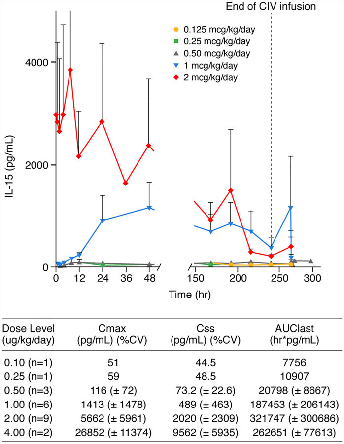
Patients and methods: Patients received treatment for 10 days with CIV rhIL15 in doses of 0.125, 0.25, 0.5, 1, 2, or 4 μg/kg/day. Correlative laboratory tests included IL15 pharmacokinetic (PK) analyses, and assessment of changes in lymphocyte subset numbers.

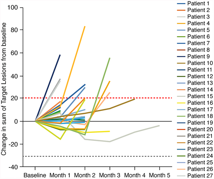
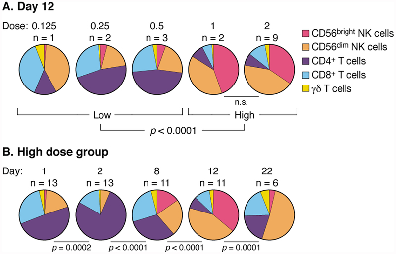
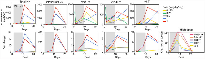
Results: Twenty-seven patients were treated with rhIL15; 2 μg/kg/day was identified as the MTD. There were eight serious adverse events including two bleeding events, papilledema, uveitis, pneumonitis, duodenal erosions, and two deaths (one due to likely drug-related gastrointestinal ischemia). Evidence of antitumor effects was observed in several patients, but stable disease was the best response noted. Patients in the 2 μg/kg/day group had a 5.8-fold increase in number of circulating CD8+ T cells, 38-fold increase in total NK cells, and 358-fold increase in CD56bright NK cells. Serum IL15 concentrations were markedly lower during the last 3 days of infusion.

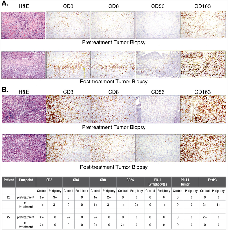
Conclusions: This phase I trial identified the MTD for CIV rhIL15 and defined a treatment regimen that produced significant expansions of CD8+ T and NK effector cells in circulation and tumor deposits. This regimen has identified several biological features, including dramatic increases in numbers of NK cells, supporting trials of IL15 with anticancer mAbs to increase antibody-dependent cell-mediated cytotoxicity and anticancer efficacy.

Trial registration: ClinicalTrials.gov NCT01572493.


Conflict of interest statement

Conflict of interest: The authors declare no potential conflicts of interest.

Figures

Figure 1.
Figure 1.. Spider Plots of Response to CIVrhIL-15 Treatment
Spider plots of all treated patients showing changes from baseline in the tumor burden (y-axis), measured as the product of the longest diameters of solid metastatic target lesions > 1 cm on high resolution CT scans (shortest diameter for lymph nodes) assessed at the end of every CIVrhIL-15 cycle (x-axis). Above the dashed red line (>20%) indicates progressive disease by RECIST criteria and below the lower black dashed line (> 30%) indicates partial response. Patients who had stable disease after their first 2 cycles of treatment continued to be restaged at regular intervals even though their treatment had been stopped.
Figure 2.
Figure 2.. Altered composition of lymphocyte subsets in response to CIV IL-15 administration
A. Representation of lymphocyte subsets at low and high dose groups after continuous infusion of rhIL-15. On day 12, two days after treatment cessation, lymphocyte representations in patients receiving low doses of IL-15 (0.125, 0.25, and 0.5 mcg/kg/day) were not different from each other but were significantly different from patients receiving 1.0 and 2.0 mcg/kg/day (p = 0.003 and p = 0.0002, respectively). Based on this, we grouped patients receiving 0.125, 0.25, and 0.5 mcg/kg/day as “low dose” and patients receiving 1.0 and 2.0 mcg/kg/day as “high dose”. These groups differed significantly at p < 0.0001. B. Kinetics of lymphocyte representations before, during, and after administration of CIV IL-15 at high dose. The relative proportion of CD56+bright NK cells was increased by day 8 of the treatment. This proportion was further increased on day 12, two days after cessation of the IL-15 infusion. By day 22, the proportions were normalizing. All statistics performed by permutation test.
Figure 3.
Figure 3.. Treatment-related changes in lymphocyte subsets
Data are shown as the mean for each dose group of CIV rhIL-15, unless otherwise stated. Fold change values were computed by individual relative to their pre-infusion baseline. Shaded gray areas indicate continuous rhIL-15 infusion. It should be noted that the vertical axes numbers differ to facilitate the analyses of the different lymphocyte populations. Absolute count and fold changes for total NK cells, CD56bright NK cells, CD8+ T cells, CD4+ T cells, and ɣδ T cells. Data for all patients are shown in Supplemental Figure S3. The frequency of cells expressing Ki-67, a marker for recent cell division, of cell subsets in the high dose group.
Figure 4.
Figure 4.. Pharmacokinetic Analysis of Mean Serum IL-15 Levels During CIV rhIL-15 Treatment
PK samples were obtained at multiple timepoints during day 1 at the end of CIV rhIL-15 (decline PK analysis) on day 11. After the 3–4 day timepoint no samples were obtained for the subsequent 3 days and single daily “steady state” samples were obtained on days 7 through 10.
Figure 5.
Figure 5.. Immunochemical Analysis of Cellular Infiltrates and Tumor Deposits
Pre-and-post-treatment analysis of the core biopsy specimen subjected to hematoxylin and eosin staining as well as immunochemical analysis for CD3 (OKT-3), CD4 (S3.5), CD8 (3B5), CD56 (56CO4), CD163 (ED2), PD-1 (MIH4) and PD-L1 (29E.2A3) was performed as previously reported (31). A. Histology from a patient with squamous cell head and neck cancer B. Histology from a patient with metastatic colorectal cancer.

References

    1. Choudhury M, Efros M, Mittelman A. Interferons and interleukins in metastatic renal cell carcinoma. Urology 1993; 41:67–72. - PubMed
    1. Kaufman HL, Kirkwood JM, Hodi FS, Agarwala S, Amatruda T, Bines SD, et al. The Society for Immunotherapy of Cancer consensus statement on tumour immunotherapy for the treatment of cutaneous melanoma. Nat Rev Clin Oncol 2013; 10:588–598. - PubMed
    1. Rosenberg SA. IL-2: the first effective immunotherapy for human cancer. J Immunol 2014; 192:5451–5458. - PMC - PubMed
    1. Linehan WM, Walther MM, Alexander RB, Rosenberg SA. Adoptive immunotherapy of renal cell carcinoma: studies from the Surgery Branch, National Cancer Institute. Semin Urol 1993; 11:41–43. - PubMed
    1. Atkins MB, Lotze MT, Dutcher JP, Fisher RI, Weiss G, Margolin K, et al. High-dose recombinant interleukin 2 therapy for patients with metastatic melanoma: analysis of 270 patients treated between 1985 and 1993. J Clin Oncol 1999; 17:2105–2116. - PubMed

Publication types

MeSH terms

Associated data