. 2019 Jul 5;294(27):10392-10406.
doi: 10.1074/jbc.RA119.007743. Epub 2019 May 29.

Defining α-synuclein species responsible for Parkinson's disease phenotypes in mice

Affiliations

Defining α-synuclein species responsible for Parkinson's disease phenotypes in mice

Jessica M Froula et al. J Biol Chem. .

Erratum in

Abstract

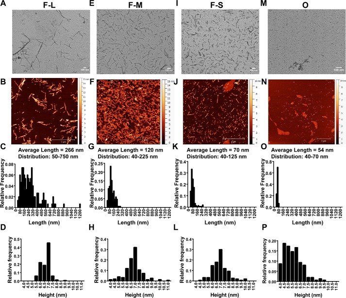
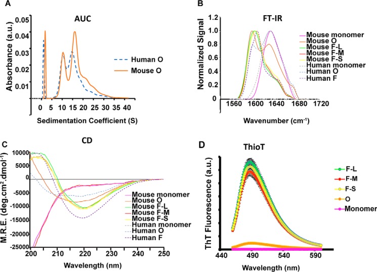
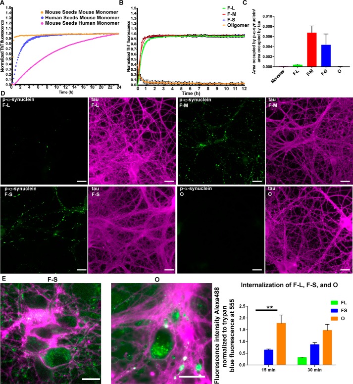
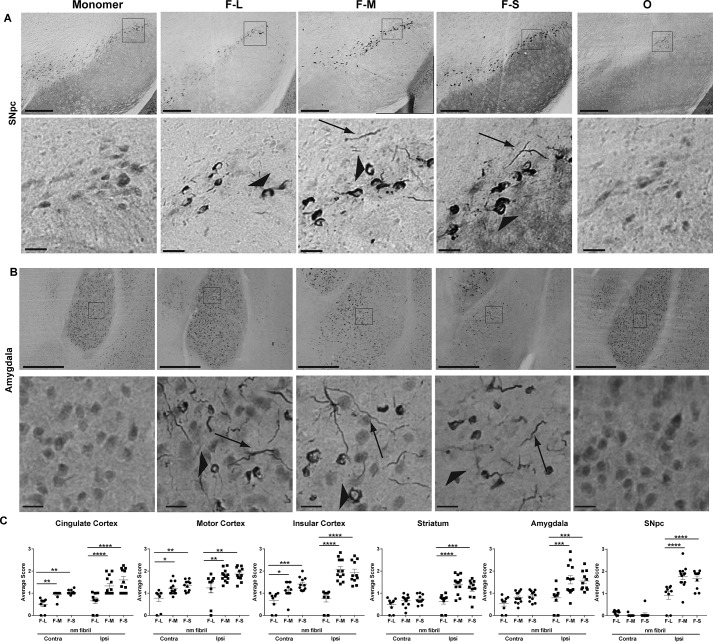
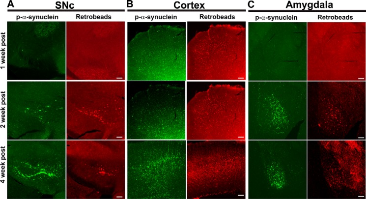
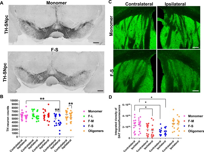
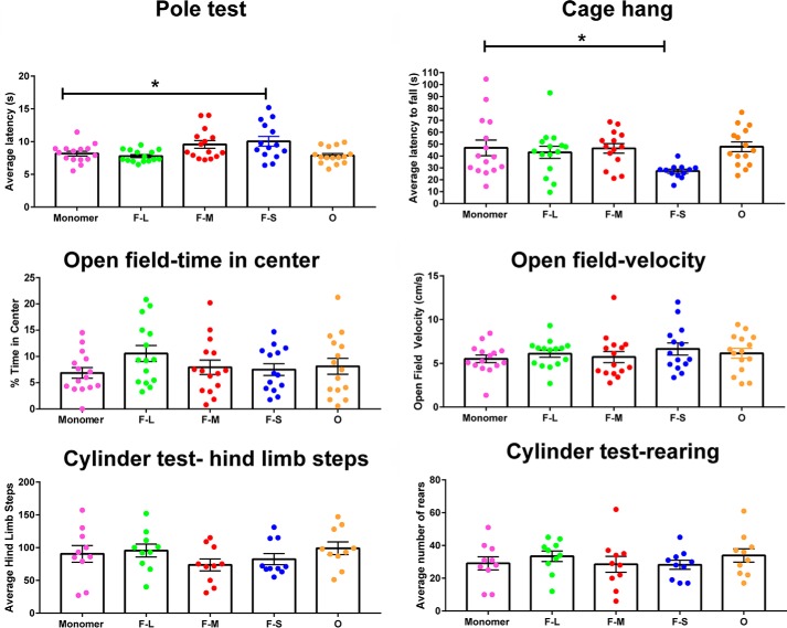
Parkinson's disease (PD) is a neurodegenerative disorder characterized by fibrillar neuronal inclusions composed of aggregated α-synuclein (α-syn). These inclusions are associated with behavioral and pathological PD phenotypes. One strategy for therapeutic interventions is to prevent the formation of these inclusions to halt disease progression. α-Synuclein exists in multiple structural forms, including disordered, nonamyloid oligomers, ordered amyloid oligomers, and fibrils. It is critical to understand which conformers contribute to specific PD phenotypes. Here, we utilized a mouse model to explore the pathological effects of stable β-amyloid-sheet oligomers compared with those of fibrillar α-synuclein. We biophysically characterized these species with transmission EM, atomic-force microscopy, CD spectroscopy, FTIR spectroscopy, analytical ultracentrifugation, and thioflavin T assays. We then injected these different α-synuclein forms into the mouse striatum to determine their ability to induce PD-related phenotypes. We found that β-sheet oligomers produce a small but significant loss of dopamine neurons in the substantia nigra pars compacta (SNc). Injection of small β-sheet fibril fragments, however, produced the most robust phenotypes, including reduction of striatal dopamine terminals, SNc loss of dopamine neurons, and motor-behavior defects. We conclude that although the β-sheet oligomers cause some toxicity, the potent effects of the short fibrillar fragments can be attributed to their ability to recruit monomeric α-synuclein and spread in vivo and hence contribute to the development of PD-like phenotypes. These results suggest that strategies to reduce the formation and propagation of β-sheet fibrillar species could be an important route for therapeutic intervention in PD and related disorders.

Keywords: Lewy body; Parkinson's disease; amyloid; cytotoxicity; fibril; motor-behavior defect; neurodegenerative disease; oligomer; protein aggregation; α-synuclein.


Conflict of interest statement

The authors declare that they have no conflicts of interest with the contents of this article

Figures

Figure 1.
Figure 1.
Morphological characterization of the mouse α-synuclein species used in the in vivo mouse studies. TEM and AFM images of fibrils (F-L) (A and B), sonicated fibrils (F-M) (E and F), sonicated fibrils enriched in short fragments (F-S) (I and J), and O (M and N). C–P, AFM was used to quantify the length and height of each species shown in the histograms.
Figure 2.
Figure 2.
Structural characterization of α-synuclein species used in the in vivo mouse studies. A, absorbance unit (a.u.) sedimentation velocity measurement of human (blue dashed line) and mouse oligomers (orange line) shows 10S and 15S species. B, FTIR spectra show that F-L, F-M, and F-S species are primarily composed of parallel β-sheets (band at 1620–1630) and that the oligomeric (mouse and human) species are primarily antiparallel (band at 1620–1630 cm−1 and shoulder at 1695 cm−1). The mouse and human oligomers have β-sheet structures of about 40 and 65%, respectively. C, CD shows that β-sheet content of oligomers (mouse and human) are intermediate between monomer and fibrils. M.R.E., mean residue ellipticity. D, ThioT binding shows that the fibrils adopt an amyloid conformation; the oligomers show limited ThioT binding, and the monomer shows no ThioT binding.
Figure 3.
Figure 3.
Seeding ability of the difference assembled forms of α-synuclein species in vitro and in primary neurons. A and B, monomer (100 μm) was incubated with 5 μm fibrillar or oligomeric seeds, and the fluorescence of samples incubated with ThioT was quantified over time. C and D, for the primary hippocampal neurons, 70 nm F-L, F-M, F-S, or oligomers were added to the neurons, and after 7 days, the neurons were fixed, and inclusion formation was visualized using an antibody to p-α-synuclein (green). Immunofluorescence for tau (magenta) shows the distribution of axons (scale bar, 50 mm). ImageJ was used to quantify the percent area occupied by p-α-synuclein fluorescence normalized to the area occupied by tau fluorescence. The data are presented as the mean ± S.E. E, primary hippocampal neurons were preincubated with Alexa488-tagged F-L, F-S or O for 30 min at 4 °C to allow binding to the cell surface. The neurons were then incubated for 15 or 30 min at 37 °C to allow internalization. Fluorescence of external α-synuclein–Alexa488 was quenched using trypan blue. Images show representative α-synuclein–Alexa488 fibrils or oligomers. When trypan blue binds to proteins on the cell surface, it fluoresces at 560 nm, which is shown in the images as magenta (scale bar, 50 μm). The fluorescence intensity of Alexa488 from 10 fields per condition was quantified and normalized to trypan blue immunofluorescence. The internalization experiments were repeated two times. **, p < 0.01.
Figure 4.
Figure 4.
Inclusion formation in the mouse brain after injection of different forms of α-synuclein. C57BL/6J mice received unilateral striatal injections of 2 μl of soluble monomer (300 μm), F-L (150 μm), F-M (150 μm), F-S (150 μm), and O (300 μm). After 3 months, mice were perfused, and immunohistochemistry was performed using an antibody to p-α-synuclein. Representative images from the SNc (A) and amygdala (B) are shown. Arrowheads indicate inclusions in the soma, and arrows indicate Lewy neurite-like inclusions. C, abundance of inclusions was measured by an investigator blinded to experimental conditions (supporting material 1). Numbers of mice are as follows: monomer (12); F-L (11); F-M (12); F-S (11); and O (15). Data are shown as the mean score ± S.E. and were analyzed using a two-way ANOVA, α-synuclein species/cingulate F(2,30) = 37.85, p < 0.0001; α-synuclein species/motor cortex F(2,30) = 7.9, p < 0.002; α-synuclein species/insular cortex F(2,30) = 22.3, p < 0.0001; α-synuclein species/striatum F(2,30) = 8.5, p < 0.001; α-synuclein species/amygdala F(2,30) = 6.6, p = 0.004; α-synuclein species/SNc F(2,30) = 6.2, p < 0.005. *, p < 0.05; **, p < 0.01; ***, p < 0.001; ****, p < 0.0001. Scale bar, 100 μm (top panels); 20 μm (bottom panels).
Figure 5.
Figure 5.
Appearance of p-α-synuclein inclusions in brain areas that project to the striatum. Fibrils and retrotracer beads were co-injected unilaterally into the striatum. After 1 week (n = 3), 2 weeks (n = 3), or 4 weeks (n = 3), mice were perfused, and immunofluorescence to p-α-synuclein (green) was performed. The retrotracer beads are shown in red, and the merged images include p-α-synuclein, retrotracer beads, and Hoechst (blue). Representative images from the SNc, motor cortex, and amygdala are shown.
Figure 6.
Figure 6.
Quantitation of TH-positive neurons in the SNc and DAT terminals in striatum following unilateral striatal injections of different α-synuclein species. C57BL/6J mice received unilateral striatal injections of 2 μl of soluble monomer (300 μm), F-L (150 μm), F-M (150 μm), F-S (150 μm), and O (300 μm). After 3 months, the mice were perfused, and immunostaining was performed. Numbers of mice are as follows: monomer (12); F-L (11); F-M (12); F-S (11); and O (15). A, representative images of tyrosine hydroxylase immunohistochemistry in the SNc of monomer- and F-S–injected mice. B, unbiased stereology of tyrosine hydroxylase–positive neurons performed by an investigator blinded to experimental conditions. Data are shown as the mean counts ± S.E. and analyzed using a two-way ANOVA, α-synuclein species F(5,67) = 2.7, p = 0.03. C, immunofluorescence performed using an antibody to DAT. Images were captured using confocal microscopy. Representative images of the striatum from monomer and F-S–injected mice are shown. D, ImageJ was used to quantify the integrated fluorescence intensity of DAT in the striatum. Data are shown as the mean counts ± S.E. and analyzed using a two-way ANOVA, α-synuclein species F(3,43) = 5.7, p = 0.002. *, p < 0.05; **, p < 0.01. Scale bar, 100 μm.
Figure 7.
Figure 7.
Motor behavior of mice following unilateral striatal injections of different α-synuclein species. C57BL/6J mice received unilateral striatal injections of 2 μl of monomer (300 μm), F-L (150 μm), F-M (150 μm), F-S (150 μm), and O (300 μm). Three months later, mice were subjected to the following behavior tests: open field, pole test, cage hang, and cylinder test (modified for mice). Numbers of mice for pole test, cage hang, and open field are as follows: monomer (12); F-L (11); F-M (12); F-S (11); and O (15). Numbers of mice for cylinder test are as follows: monomer (10); F-L (10); F-M (10); F-S (10); and O (10). The data were analyzed by one-way ANOVA: pole test F(4,69) = 4.7, p = 0.002; cage hang F(4,66) = 2.86, p = 0.03; open field/% time center F(4,69) = 1,1, p = not significant; open field/velocity F(4,69) = 0.6, p = not significant; hind limb steps F(4,45) = 1.1, p = not significant; rearing F(4,45) = 0.5, p = not significant. *, p < 0.05.

References

    1. Cremades N., and Dobson C. M. (2018) The contribution of biophysical and structural studies of protein self-assembly to the design of therapeutic strategies for amyloid diseases. Neurobiol. Dis. 109, 178–190 10.1016/j.nbd.2017.07.009 - DOI - PubMed
    1. Spillantini M. G., Schmidt M. L., Lee V. M., Trojanowski J. Q., Jakes R., and Goedert M. (1997) α-Synuclein in Lewy bodies. Nature 388, 839–840 10.1038/42166 - DOI - PubMed
    1. Kuusisto E., Parkkinen L., and Alafuzoff I. (2003) Morphogenesis of Lewy bodies: dissimilar incorporation of α-synuclein, ubiquitin, and p62. J. Neuropathol. Exp. Neurol. 62, 1241–1253 10.1093/jnen/62.12.1241 - DOI - PubMed
    1. Volpicelli-Daley L. A., Luk K. C., Patel T. P., Tanik S. A., Riddle D. M., Stieber A., Meaney D. F., Trojanowski J. Q., and Lee V. M. (2011) Exogenous α-synuclein fibrils induce Lewy body pathology leading to synaptic dysfunction and neuron death. Neuron 72, 57–71 10.1016/j.neuron.2011.08.033 - DOI - PMC - PubMed
    1. Luk K. C., Kehm V., Carroll J., Zhang B., O'Brien P., Trojanowski J. Q., and Lee V. M. (2012) Pathological α-synuclein transmission initiates Parkinson-like neurodegeneration in nontransgenic mice. Science 338, 949–953 10.1126/science.1227157 - DOI - PMC - PubMed

Publication types