Review
. 2019 Jun 11;92(24):1146-1156.
doi: 10.1212/WNL.0000000000007654. Epub 2019 May 29.

CNS small vessel disease: A clinical review

Affiliations
Review

CNS small vessel disease: A clinical review

Rocco J Cannistraro et al. Neurology. .

Abstract

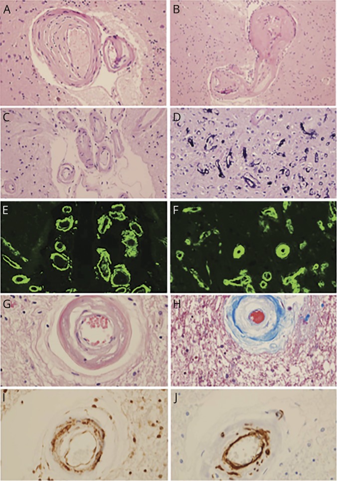
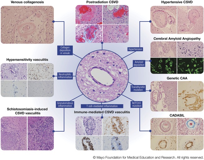
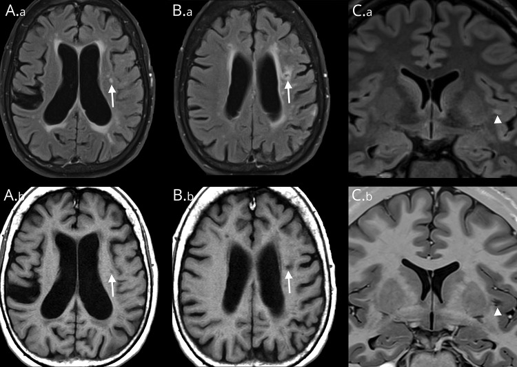
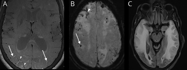
CNS small vessel disease (CSVD) causes 25% of strokes and contributes to 45% of dementia cases. Prevalence increases with age, affecting about 5% of people aged 50 years to almost 100% of people older than 90 years. Known causes and risk factors include age, hypertension, branch atheromatous disease, cerebral amyloid angiopathy, radiation exposure, immune-mediated vasculitides, certain infections, and several genetic diseases. CSVD can be asymptomatic; however, depending on location, lesions can cause mild cognitive dysfunction, dementia, mood disorders, motor and gait dysfunction, and urinary incontinence. CSVD is diagnosed on the basis of brain imaging biomarkers, including recent small subcortical infarcts, white matter hyperintensities, lacunes, cerebral microbleeds, enlarged perivascular spaces, and cerebral atrophy. Advanced imaging modalities can detect signs of disease even earlier than current standard imaging techniques. Diffusion tensor imaging can identify altered white matter connectivity, and blood oxygenation level-dependent imaging can identify decreased vascular reactivity. Pathogenesis is thought to begin with an etiologically specific insult, with or without genetic predisposition, which results in dysfunction of the neurovascular unit. Uncertainties regarding pathogenesis have delayed development of effective treatment. The most widely accepted approach to treatment is to intensively control well-established vascular risk factors, of which hypertension is the most important. With better understanding of pathogenesis, specific therapies may emerge. Early identification of pathologic characteristics with advanced imaging provides an opportunity to forestall progression before emergence of symptoms.


Figures

Figure 1
Figure 1. The most common pathologic characteristics of CNS small vessel disease
(A, B) Hypertensive CSVD (H&E stain, original magnification ×400), as evidenced by hyaline arteriolar sclerosis (A) and a microaneurysm (B). (C, D) CAA (H&E stain, original magnification ×200), as evidenced by amyloid-β-laden vessels in the subarachnoid space, with the double-barreled appearance (C) and capillaries with calcium mineralization (D) (H&E stain, original magnification ×400). (E) Leptomeningeal CAA (thioflavin S, original magnification ×200). (F) Parenchymal CAA (thioflavin S, original magnification ×400). (G) CADASIL showing loss of smooth muscle cells (H&E stain, original magnification ×400). (H) CADASIL showing collagen deposition and loss of smooth muscle cells (trichrome stain, original magnification ×400). (I) Ubiquitinated proteins in granular osmiophilic material (ubiquitin immunohistochemistry, original magnification ×400). (J) Smooth muscle cell degradation due to CADASIL (smooth muscle actin immunohistochemistry, original magnification ×400). CAA = cerebral amyloid angiopathy; CADASIL = cerebral autosomal dominant arteriopathy with subcortical infarcts and leukoencephalopathy; CSVD = CNS small vessel disease; H&E = hematoxylin-eosin.
Figure 2
Figure 2. Various pathologic characteristics of CNS small vessel disease
Left column, venous collagenosis. H&E stain of a periventricular venule in the white matter with thickened walls and collagen deposition. Hypersensitivity vasculitis. (Top left) H&E stain showing features of leukoctyoclastic vasculitis; (top right) CD68 immunohistochemistry for macrophages; (bottom left) CD20 showing sparse B lymphocytes; (bottom right) CD3 showing rare T lymphocytes. Schistosomiasis-induced CSVD vasculitis. (Left and right) H&E stains showing granulomatous inflammation. Middle column, postradiation CSVD. (Top left) H&E stain showing fibrinoid necrosis; (top right) H&E stain showing capillary ectasia and atypical nuclear changes; (bottom left) trichrome stain showing collagenosis (blue) and fibrinoid material; (bottom right) H&E stain showing obliterative collagenosis of small vessels with nuclear atypia. Center image, H&E stain showing a normal subcortical white matter arteriole. Immune-mediated CSVD vasculitis. (Top left) H&E stain showing cellular infiltrates in vessel wall; (top right) HLA-DR immunohistochemistry showing macrophage infiltrates; (bottom left) CD20 immunohistochemistry showing B-lymphocytic infiltrates; (bottom right) CD3 showing T-lymphocytic infiltrates. Right column, hypertensive CSVD. (Left) H&E stain showing hyaline arteriolar sclerosis; (right) H&E stain showing a microaneurysm. Cerebral Amyloid Angiopathy. (Top left) H&E stain showing amyloid-β-laden vessels in the subarachnoid spacewith the doublebarreled appearance; (top right) capillaries with calcium mineralization; (bottom left) leptomeningeal CAA on thioflavin S fluorescent microscopy; (bottom right) parenchymal CAA on thioflavin S fluorescent microscopy. Genetic CAA/novel transthyretin mutation. (Left) H&E stain showing leptomeningeal arteriole involvement; (right) transthyretin immunohistochemistry. CADASIL. (Top left) H&E stain showing loss of smooth muscle cells; (top right) trichrome stain showing collagen deposition (blue) in wall of affected arteriole; (bottom left) ubiquitin immunohistochemistry showing granular deposits in arterial wall; (bottom right) smooth muscle actin immunohistochemistry showing fragmentation and loss of smooth muscle cells in affected arteriole. Abbreviations: CAA = cerebral amyloid angiopathy; CADASIL = cerebral autosomal dominant arteriopathy with subcortical infarcts and leukoencephalopathy; CSVD = central nervous systemsmall vessel disease. H&E = hematoxylin-eosin (Figure used with permission of Mayo Foundation for Medical Education and Research).
Figure 3
Figure 3. MRI findings of CNS small vessel disease
Patient 1, images A.a, A.b, B.a, B.b. (A.a) Axial T2 FLAIR image shows a typical WMH (arrow) with increased FLAIR signal. (A.b) Corresponding intermediate-signal intensity (arrow) on T1-weighted MRI. (B.a) By contrast, an axial T2 FLAIR image taken slightly more superior in location shows a lacune with marginal increased FLAIR hyperintensity reflecting gliosis with central hypointensity (arrow) representing cavitation. (B.b) The corresponding axial T1-weighted image shows marginal intermediate signal (arrow) corresponding to areas of gliosis with central hypointensity indicating cavitation. Patient 2, images C.a and C.b. (C.a) Coronal T2 FLAIR image shows a left-sided FLAIR hyperintensity (arrowhead) that represents incomplete fluid suppression within a PVS, which is a common diagnostic pitfall and may be erroneously interpreted as a WMH. (C.b) Corresponding coronal T1-weighted image better illustrates the linear branching configuration (arrowhead) typical of a PVS. FLAIR = fluid-attenuated inversion recovery; PVS = perivascular space; WMH = white matter hyperintensity.
Figure 4
Figure 4. MRI findings characteristic of cerebral amyloid angiopathy
(A) Axial postcontrast vessel wall image showing enhancement of the posterior vessel walls (arrows) with multiple chronic perivascular microhemorrhages (arrowheads). (B) Susceptibility-weighted image showing numerous chronic peripheral CMBs (arrow) and cSS (arrowhead) in a patient with CAA. The microhemorrhages show “blooming artifact” in which the lesions appear larger than their actual sizes because of a magnetic susceptibility effect. (C) T2-weighted FLAIR sequence showing a confluent hyperintensity that is consistent with vasogenic edema from CAA–related inflammation. CAA = cerebral amyloid angiopathy; CMB = cerebral microbleed; cSS = cortical superficial siderosis; FLAIR = fluid-attenuated inversion recovery.

References

    1. Nam KW, Kwon HM, Lim JS, Han MK, Nam H, Lee YS. The presence and severity of cerebral small vessel disease increases the frequency of stroke in a cohort of patients with large artery occlusive disease. PLoS One 2017;12:e0184944. - PMC - PubMed
    1. Gorelick PB, Scuteri A, Black SE, et al. . Vascular contributions to cognitive impairment and dementia: a statement for healthcare professionals from the American Heart Association/American Stroke Association. Stroke 2011;42:2672–2713. - PMC - PubMed
    1. Inzitari D, Pracucci G, Poggesi A, et al. . Changes in white matter as determinant of global functional decline in older independent outpatients: three year follow-up of LADIS (leukoaraiosis and disability) study cohort. BMJ 2009;339:b2477. - PMC - PubMed
    1. Fisher CM. Lacunar strokes and infarcts: a review. Neurology 1982;32:871–876. - PubMed
    1. Pantoni L. Cerebral small vessel disease: from pathogenesis and clinical characteristics to therapeutic challenges. Lancet Neurol 2010;9:689–701. - PubMed

MeSH terms

Substances