. 2019 May 29;11(494):eaao0498.
doi: 10.1126/scitranslmed.aao0498.

Sustained correction of associative learning deficits after brief, early treatment in a rat model of Fragile X Syndrome

Affiliations

Sustained correction of associative learning deficits after brief, early treatment in a rat model of Fragile X Syndrome

Antonis Asiminas et al. Sci Transl Med. .

Abstract

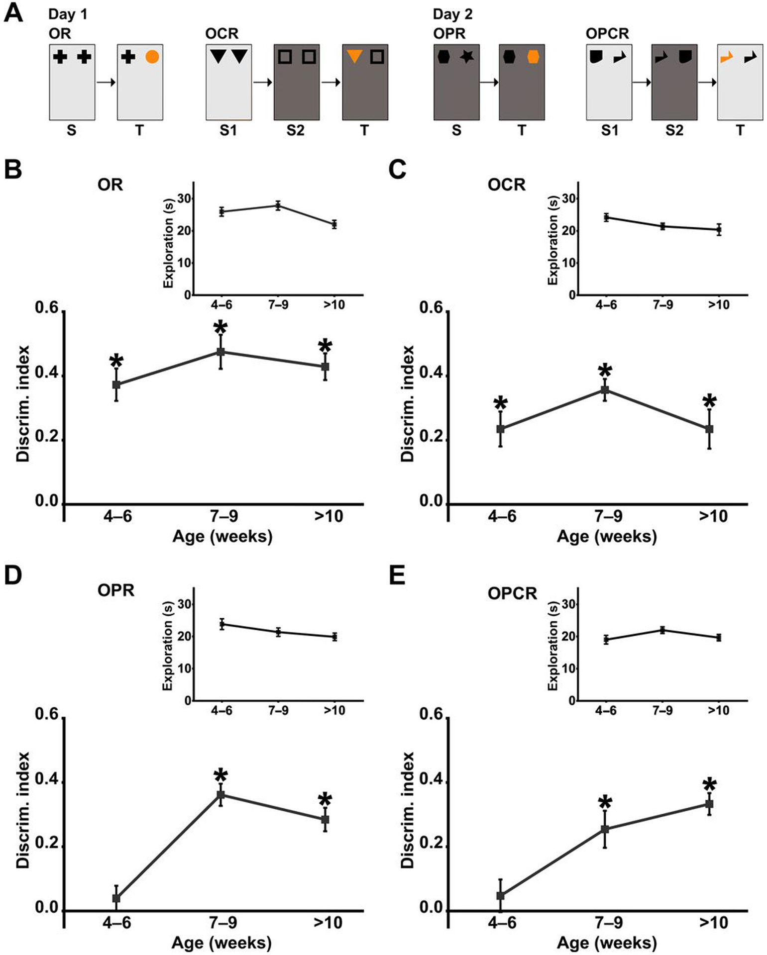
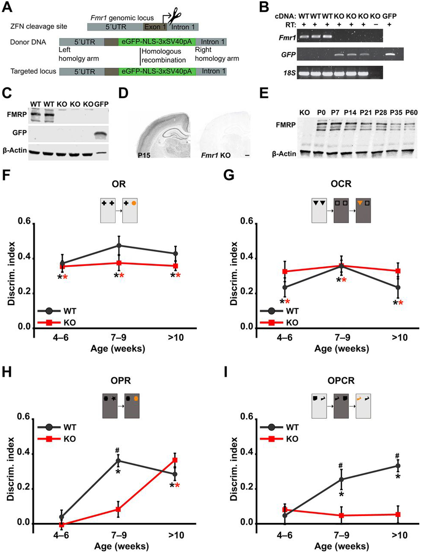
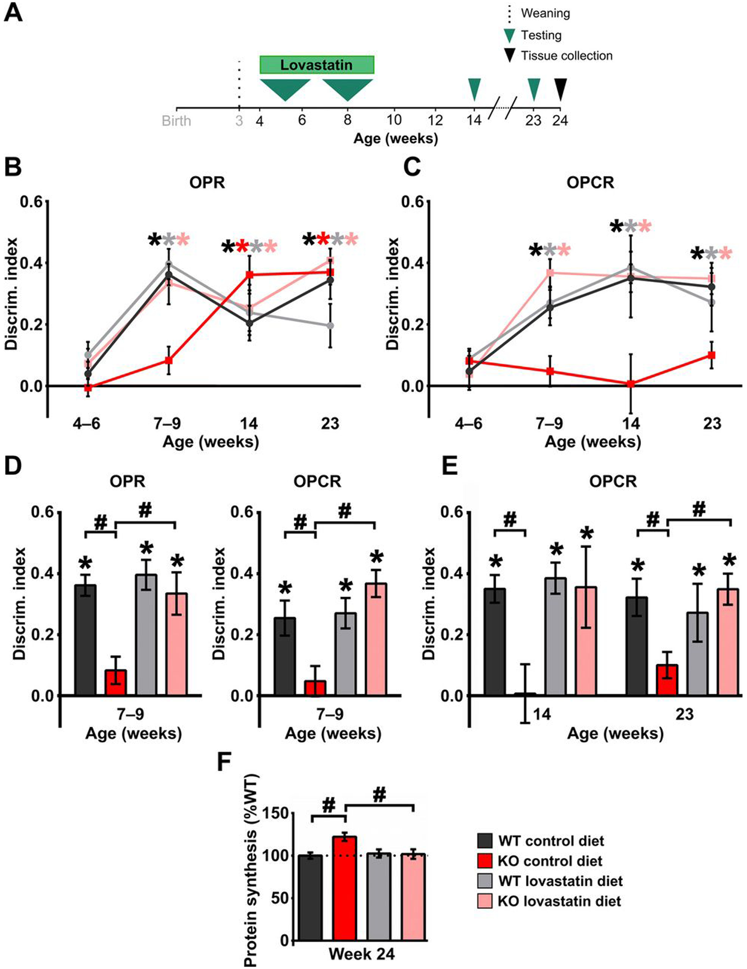
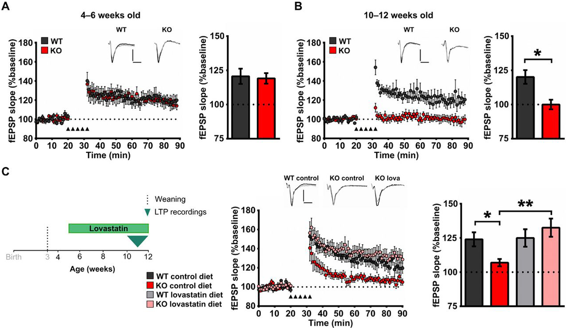
Fragile X Syndrome (FXS) is one of the most common monogenic forms of autism and intellectual disability. Preclinical studies in animal models have highlighted the potential of pharmaceutical intervention strategies for alleviating the symptoms of FXS. However, whether treatment strategies can be tailored to developmental time windows that define the emergence of particular phenotypes is unknown. Similarly, whether a brief, early intervention can have long-lasting beneficial effects, even after treatment cessation, is also unknown. To address these questions, we first examined the developmental profile for the acquisition of associative learning in a rat model of FXS. Associative memory was tested using a range of behavioral paradigms that rely on an animal's innate tendency to explore novelty. Fmr1 knockout (KO) rats showed a developmental delay in their acquisition of object-place recognition and did not demonstrate object-place-context recognition paradigm at any age tested (up to 23 weeks of age). Treatment of Fmr1 KO rats with lovastatin between 5 and 9 weeks of age, during the normal developmental period that this associative memory capability is established, prevents the emergence of deficits but has no effect in wild-type animals. Moreover, we observe no regression of cognitive performance in the FXS rats over several months after treatment. This restoration of the normal developmental trajectory of cognitive function is associated with the sustained rescue of both synaptic plasticity and altered protein synthesis. The findings provide proof of concept that the impaired emergence of the cognitive repertoire in neurodevelopmental disorders may be prevented by brief, early pharmacological intervention.


Conflict of interest statement

Competing interests: M.F.B. holds a patent titled “Methods of treating disorders with group I mGluR antagonists” (US6890931B2) and has served as a paid consultant to Q-State Biosciences, Vertex Pharmaceuticals, and Sunovion Pharmaceuticals.

Figures

Fig. 1.
Fig. 1.. WT LEH rats show distinct developmental trajectories in different spontaneous object exploration tasks.
(A) Schematic of the spontaneous object exploration tasks used. S, sample phase; T, test phase. Light and dark gray backgrounds denote distinct contexts, and orange symbols denote novel object/object association. Discrimination index (DI) over time for WT LEH rats in (B) OR, (C) OCR, (D) OPR, and (E) OPCR tasks. Insets (B to E) Total object exploration time during the test phase for each task. For all tasks, n(4–6) = 13, n(7–9) = 13, n(>10) = 11; *P < 0.05 difference from chance (DI = 0) based on one-sample t tests. P values from one-sample t tests have been corrected for false discovery rate using the Benjamini-Hochberg procedure. For details on t, df, and P values for one-sample t tests, see table S1.
Fig. 2.
Fig. 2.. Loss of FMRP leads to selective deficits in OPR and OPCR memory in Fmr1 KO rats.
(A) Zinc finger nuclease (ZFN)–mediated disruption of Fmr1. Diagrams illustrate the target site for ZFN cleavage, donor DNA sequence including enhanced green fluorescent protein (eGFP) and a nuclear localization signal flanked by 5′ and 3′ homology recombination arms for homology-directed repair, and the resulting targeted locus (top). 5′UTR, 5′ untranslated region. (B) RT-PCR for Fmr1 and eGFP mRNA in WT and Fmr1 KO rats. Lanes 1 to 3, samples from three WT rats; lanes 4 to 6, samples from three Fmr1 KO rats; lane 7, RT control from one Fmr1 KO rat; lane 8, GFP-positive control. cDNA, complementary DNA. (C) Western blot analyses of FMRP and GFP expression in WT and Fmr1 KO rats. Lanes 1 to 2, samples from two WT rats; lanes 3 to 5, samples from three Fmr1 KO rats; lane 6, positive control for GFP. (D) Immunohistochemical localization of FMRP in P15 WT and Fmr1 KO rats. Scale bar, 500 μm. (E) Western blot analysis of FMRP expression in hippocampal homogenates from WT littermates over postnatal development compared with P15 Fmr1 KO rat. DI at different ages for WT and Fmr1 KO rats in (F) OR, (G) OCR, (H) OPR, and (I) OPCR tasks. For all tasks, nWT(4–6) = 13, nWT(7–9) = 13, nWT(>10) = 11, nKO(4–6) = 12, nKO(7–9) = 12, nKO(>10) = 11; *P< 0.05 difference from chance (DI = 0), black for WT and red for KO; #P< 0.05 difference between groups. Linear mixed effect (LME) models were fitted to the data (for details, see tables S7 and S8). P values from one-sample t tests and post hoc two-sample t tests have been controlled for the false discovery rate using the Benjamini-Hochberg procedure. For details on t, df, and P values for one-sample t tests and post hoc two-sample t tests, see table S1.
Fig. 3.
Fig. 3.. Transient treatment with lovastatin restores WT-like developmental trajectory of OPR and OPCR memory in Fmr1 KO rats and has sustained effects on both memory and cellular pathophysiology.
(A) Experimental time course for longitudinal assessment of cognitive development in WT and Fmr1 KO rats treated with lovastatin between 4 and 9 weeks of age. DI at different ages for WT and Fmr1 KO rats treated with control or lovastatin diet in (B) OPR and (C) OPCR tasks. (D) Effect of lovastatin treatment on OPR (left) and OPCR (right) memory tested at 7 to 9 weeks of age in WT and Fmr1 KO rats. (E) Effect of lovastatin treatment on OPCR memory in WT and Fmr1 KO rats tested at 14 and 23 weeks of age. (F) Effect of lovastatin treatment on hippocampal basal protein synthesis levels in WT and Fmr1 KO rats measured at 24 weeks of age, after behavioral testing was complete. Sample sizes: OPR 4 to 6 weeks: nWTcontrol = 13, nWTlova = 12, nKOcontrol = 12, nKOlova = 12; OPR 7 to 9 weeks: nWTcontrol = 13, nWTlova = 12, nKOcontrol = 12, nKOlova = 12; OPR 14 weeks: nWTcontrol = 10, nWTlova = 11, nKOcontrol = 11, nKOlova = 8; OPR 23 weeks: nWTcontrol = 11, nWTlova = 10, nKOcontrol = 11, nKOlova = 7; OPCR 4 to 6 weeks: nWTcontrol = 13, nWTlova = 12, nKOcontrol = 12, nKOlova = 12; OPCR 7 to 9 weeks: nWTcontrol = 13, nWTlova = 12, nKOcontrol = 12, nKOlova = 12; OPCR 14 weeks: nWTcontrol = 11, nWTlova = 11, nKOcontrol = 11, nKOlova = 8; OPCR 23 weeks: nWTcontrol = 10, nWTlova = 10, nKOcontrol = 11, nKOlova = 8; for protein synthesis, n = 6 for all groups. *P< 0.05 difference from chance (DI = 0), black for WT and red for KO; #P< 0.05 difference between groups. LMEs were fitted to the behavioral data (for details, see tables S7 and S9), and two-way analysis of variance (ANOVA) with post hoc two-sample t tests was used to analyze the effect of lovastatin on hippocampal protein synthesis levels (for details, see table S6F). P values from one-sample t tests and post hoc two-sample t tests have been controlled for the false discovery rate using the Benjamini-Hochberg procedure. For details on behavioral data t, df, and P values for one-sample t tests, see table S3, and for post hoc two-sample t tests, see table S4.
Fig. 4.
Fig. 4.. Lovastatin prevents the emergence of plasticity deficits associated with the loss of FMRP.
(A) Left: Time course plotting field excitatory postsynaptic potential (fEPSP) slopes normalized to baseline after LTP induction in layers 2 to 5 synapses in the prelimbic mPFC taken from 4- to 6-week-old WT and Fmr1 KO rats. Right: Averages of fEPSP slopes normalized to baseline during the last 20 min of the recording (70 to 90 min). (B) Left: Time course plotting fEPSP slopes normalized to baseline after LTP induction at synapses from layer 2/3 inputs onto layer 5 neurons in the prelimbic mPFC taken from 10- to 12-week-old WT and Fmr1 KO rats. Right: Averages of fEPSP slopes normalized to baseline during the last 20 min of the recording (70 to 90 min). (C) Left: Experimental time course for assessment of the effect of lovastatin (lova) treatment beginning at 5 weeks of age on WT and Fmr1 KO plasticity in the prelimbic mPFC. Middle: Time course plotting averages of fEPSP slopes normalized to baseline after LTP induction in layers 2 to 5 synapses in prelimbic mPFC slices taken from 10- to 12-week-old WT and Fmr1 KO treated with either control or lovastatin diet. Right: Averages of fEPSP slopes normalized to baseline during the last 20 min of recordings (70 to 90 min). Insets: Example traces showing synaptic responses during baseline (black trace) and at 80 to 90 min (gray trace). Scale bar, 0.5 mV; 5 ms. Sample sizes: LTP 4 to 6 weeks, n = 6 for WT and KO; LTP 10 to 12 weeks, n = 7 for WT and KO; for lovastatin effects on LTP, nWTcontrol = 9, nWTlova = 7, nKOcontrol = 9, nKOlova = 8; *P < 0.05; **P < 0.01 difference between groups; two-way ANOVA with post hoc two-sample t tests was used for data analyses (for details, see table S6, G and H). P values for post hoc two-sample t tests have been controlled for the false discovery rate using the Benjamini-Hochberg procedure.

References

    1. Kaufmann WE, Kidd SA, Andrews HF, Budimirovic DB, Esler A, Haas-Givler B, Stackhouse T, Riley C, Peacock G, Sherman SL, Brown WT, Berry-Kravis E, Autism spectrum disorder in fragile X syndrome: Cooccurring conditions and current treatment. Pediatrics 139, S194–S206 (2017). - PMC - PubMed
    1. Hagerman RJ, Berry-Kravis E, Hazlett HC, Bailey DB Jr., Moine H, Kooy RF, Tassone F, Gantois I, Sonenberg N, Mandel JL, Hagerman PJ, Fragile X syndrome. Nat. Rev. Dis. Primers 3, 17065 (2017). - PubMed
    1. Harlow EG, Till SM, Russell TA, Wijetunge LS, Kind P, Contractor A, Critical period plasticity is disrupted in the barrel cortex of Fmr1 knockout mice. Neuron 65, 385–398 (2010). - PMC - PubMed
    1. He Q, Nomura T, Xu J, Contractor A, The developmental switch in GABA polarity is delayed in fragile X mice. J. Neurosci 34, 446–450 (2014). - PMC - PubMed
    1. Verkerk AJMH, Pieretti M, Sutcliffe JS, Fu Y-H, Kuhl DPA, Pizzuti A, Reiner O, Richards S, Victoria MF, Zhang F, Eussen BE, van Ommen G-JB, Blonden LAJ, Riggins GJ, Chastain JL, Kunst CB, Galjaard H, Thomas Caskey C, Nelson DL, Oostra BA, Warran ST, Identification of a gene (FMR-1) containing a CGG repeat coincident with a breakpoint cluster region exhibiting length variation in fragile X syndrome. Cell 65, 905–914 (1991). - PubMed

Publication types

MeSH terms

Substances