. 2019 Jun;570(7760):241-245.
doi: 10.1038/s41586-019-1257-5. Epub 2019 May 29.

Cas13-induced cellular dormancy prevents the rise of CRISPR-resistant bacteriophage

Affiliations

Cas13-induced cellular dormancy prevents the rise of CRISPR-resistant bacteriophage

Alexander J Meeske et al. Nature. 2019 Jun.

Abstract

Clustered, regularly interspaced, short palindromic repeat (CRISPR) loci in prokaryotes are composed of 30-40-base-pair repeats separated by equally short sequences of plasmid and bacteriophage origin known as spacers1-3. These loci are transcribed and processed into short CRISPR RNAs (crRNAs) that are used as guides by CRISPR-associated (Cas) nucleases to recognize and destroy complementary sequences (known as protospacers) in foreign nucleic acids4,5. In contrast to most Cas nucleases, which destroy invader DNA4-7, the type VI effector nuclease Cas13 uses RNA guides to locate complementary transcripts and catalyse both sequence-specific cis- and non-specific trans-RNA cleavage8. Although it has been hypothesized that Cas13 naturally defends against RNA phages8, type VI spacer sequences have exclusively been found to match the genomes of double-stranded DNA phages9,10, suggesting that Cas13 can provide immunity against these invaders. However, whether and how Cas13 uses its cis- and/or trans-RNA cleavage activities to defend against double-stranded DNA phages is not understood. Here we show that trans-cleavage of transcripts halts the growth of the host cell and is sufficient to abort the infectious cycle. This depletes the phage population and provides herd immunity to uninfected bacteria. Phages that harbour target mutations, which easily evade DNA-targeting CRISPR systems11-13, are also neutralized when Cas13 is activated by wild-type phages. Thus, by acting on the host rather than directly targeting the virus, type VI CRISPR systems not only provide robust defence against DNA phages but also prevent outbreaks of CRISPR-resistant phage.


Conflict of interest statement

Competing interests: L.A.M. is a cofounder and Scientific Advisory Board member of Intellia Therapeutics, and a co-founder of Eligo Biosciences.

Figures

Extended Data Fig. 1.
Extended Data Fig. 1.. Listeria phage infection model for studying type VI-A CRISPR immunity.
(a) Diagram of the ϕRR4 genome, with individual genes depicted within the anti-CRISPR, early lytic, late lytic, and lysogenic regions. Listeria seeligeri ATCC35967 harbors a five-spacer type VI-A CRISPR locus, but phages infecting this strain have not yet been identified. We sequenced the genome of L. seeligeri RR4 and found it contains a 42 kb prophage, ϕRR4, that is similar to the A118 listeriophage. Although ϕRR4 particles induced from the lysogen did not infect L. seeligeri ATCC35967, ϕRR4 propagated in the closely related Listeria ivanovii RR3 strain (99.2% 16S rRNA identity). (b) The type VI-A CRISPR locus of L. seeligeri ATCC35967 was inserted into the tRNAArg gene of Listeria ivanovii RR3 using the vector pAM125, generating L. ivanovii ΩCRISPRVI. Different strains with either the five spacers naturally present in this system (spc1-5), individual spacers matching the genome of ϕRR4 (spcA, spcE, spcL), or a ϕRR4 spacer library (spc lib), were generated. For the latter, 41,276 ϕRR4-matching spacers were selected, tiled every 2 nt across the phage genome, with both strands equally represented. (c) Test of type VI-A anti-plasmid immunity in L. ivanovii ΩCRISPRVI(spc1-5). Plasmids with spc2 or spc4 targets in the chloramphenicol resistance cassette were conjugated into L. ivanovii RR3 or ΩCRISPRVI(spc1-5) and transconjugants selected on nalidixic acid and chlorampehnicol. Transconjugants receiving an empty vector lacking a target sequence are shown as a negative control. Example is representative of two biological replicates. (d) Prevention of ϕRR4 lytic infection by the type VI spacer library. Spacer library cells (yellow-orange gradient) or cells lacking CRISPR (gray) were infected with ϕRR4 at OD600 = 0.1, MOI=1 and OD600 was monitored over time. Example is representative of two biological replicates. (e) 100 bp sliding window average spacer enrichment ratio (spacer abundance post-infection / pre-infection) for spacers targeting top (orange) and bottom (brown) strands.
Extended Data Fig. 2.
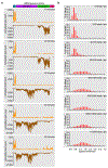
Extended Data Fig. 2.. ϕRR4 transcriptome and enrichment of corresponding targeting spacers.
(a) RNA-seq over the course of ϕRR4 infection. Wild-type L. ivanovii RR3 was infected with ϕRR4 at MOI 1 and samples were harvested for transcriptomic analysis by paired-end RNA-seq at the indicated time points. Reads were mapped to the ϕRR4 genome and normalized to the total reads per sample. Top strand mapped reads are shown in orange, bottom strand in brown. Examples are representative of two biological replicates. (b) Spacer enrichment correlates with target transcription, with no additional protection conferred above a critical expression threshold. Spacer abundance in the library was assessed pre-infection as well as 5 hours post-infection with ϕRR4 at MOI 1. Spacer enrichment distributions are shown, with individual histograms representing different tiers of target transcript abundance for the corresponding protospacer.
Extended Data Fig. 3.
Extended Data Fig. 3.. Cas13a-mediated cleavage of phage and host transcripts detected by RNA-seq.
(a) Abundance of phage transcripts assessed by conventional paired-end RNA-seq 1.75 hours post infection with ϕRR4 in L. ivanovii RR3 wild-type, ΩCRISPRVI(spcE) or ΩCRISPRVI(spcL) strains. Reads were mapped to the ϕRR4 genome and normalized to the abundance of a spike-in RNA, with top strand reads shown in orange and bottom strand reads in brown. Both ΩCRISPRVI(spcE) and ΩCRISPRVI(spcL) targeting result in elevated early transcript cleavage products (see Fig. 2a–b) and the reduction of late transcript abundance. Examples are representative of two biological replicates. (b) L. ivanovii host mRNA cleavage detected by 5’ end mapping in L. ivanovii RR3 wild-type (gray) and ΩCRISPRVI(spcE) (green) strains 1.75 hours post infection with ϕRR4. The height of each peak represents the detected abundance of the corresponding mRNA 5’ end, with transcriptional start sites depicted as gray arrows. Four regions of the genome are depicted: murA1, ftsEX/iap, isdCD, division and cell wall (dcw) cluster. Abundant intragenic cleavage products are generated in the ΩCRISPRVI(spcE) strain. Examples are representative of two biological replicates. (c) The four corresponding genomic regions in (b) shown for the native type VI CRISPR host L. seeligeri, wild type (red) and ∆CRISPR (gray), 15 minutes after aTc-induction of a target transcript. The dcw cluster is broken into two operons in L. seeligeri. Examples are representative of two biological replicates.
Extended Data Fig. 4.
Extended Data Fig. 4.. Trans-RNase activity is sufficient to limit growth of both ϕRR4 phage and ΩCRISPRIV host.
(a) L. ivanovii RR3, ΩCRISPRVI(spcA, spcE, or spcL) strains at OD600=0.05 were infected with ϕRR4 at MOI 1 and growth was monitored over 24 hours. Each curve represents the mean of three biological replicates ±s.e.m. (b) Quantitation of ϕRR4 infective centers over time on wild-type L. ivanovii RR3. Cells were infected with ϕRR4 at MOI 0.1 and allowed to adsorb 5 minutes, then cells were washed 3 times to remove free phage. Infective centers were counted every 30 minutes by enumerating plaque-forming units on a lawn of phage-susceptible RR3 cells. Each data point represents the mean of three biological replicates ±s.e.m. (c) Survival of the indicated strains during ϕRR4 infection at MOI 2. CFU were titered pre-infection (P) and 4 hours post-infection (IN) or mock-infection (UN). Each bar represents the mean of three biological replicates ±s.e.m.
Extended Data Fig. 5.
Extended Data Fig. 5.. Cas13a activation induces reversible dormancy of the host.
(a) Growth arrest (measured as the culture OD600) induced by target transcription in wil-type L. seeligeri (but not the ∆CRISPR mutant) harboring an aTc-inducible protospacer RNA. Arrow indicates time of 100 ng/mL aTc addition. Each data point represents the mean of three biological replicates ±s.e.m. (b) Wild-type and ∆CRISPR L. seeligeri cultures carrying an aTc-inducible target transcript were exposed to 100 ng/mL aTc for 3 hours as in (a), then diluted (at time 0 hours) to OD600=0.05 in fresh media in the presence or absence of aTc, and growth was monitored over 24 hours. Each curve represents the mean of three biological replicates ±s.e.m. (c) Immediate reduction in CFU upon phage infection of L. ivanovii RR3 or ΩCRISPRVI strains. The indicated strains were infected with ϕRR4 at MOI 2, and CFU titers in the infected cultures were monitored over time. Pre-infection (P) and mock-infection titers were also measured. Each bar represents the mean of three biological replicates ±s.e.m. (d) Cell vitality within ΩCRISPRVI cultures during phage infection. Cell vitality was measured in samples of cultures from (c) by monitoring conversion of nonfluorescent resazurin to fluorescent resorufin at each time point. Resorufin signal from heat-killed cells was subtracted from all samples as background, and each signal was normalized to pre-infection value. 10% and 50% live cell standards (mixed with heat-killed cells) are shown to demonstrate quantitative capability of vitality assay. Each bar represents the mean of three biological replicates ±s.e.m. (e) Phage-susceptible L. ivanovii ΩCRISPRVI(spcP) cells harboring a spacer against an aTc-inducible plasmid target RNA (or empty vector control) were treated with aTc for 1h to pre-activate Cas13a, then infected with ϕRR4 at MOI 1. Viable CFU were enumerated pre-infection (PRE), 7 hours post-infection (T7) or mock-infection (UN). *** indicates a significant difference, determined by two-sided Student’s t-test, p=0.0005. Each bar represents the mean of three biological replicates ±s.e.m.
Extended Data Fig. 6.
Extended Data Fig. 6.. Absence of CRISPR-resistant escape mutants and validation of engineered escaper phage.
(a) Efficiency of plaquing assays with wild-type ϕRR4 and engineered spcA-escaper phage ϕRR4acr infecting L. ivanovii RR3 and ΩCRISPRVI(spcA) strains. Phages were diluted and spotted onto top agar lawns containing the indicated strain. Escaper plaques are not observed in the presence of type VI CRISPR targeting. The ϕRR4acr mutant, lacking the acr region targeted by spcA, is viable and evades CRISPR targeting. Examples are representative of two biological replicates. (b) Same as (a) but testing spcE-escaper phage ϕRR4early and the ΩCRISPRVI(spcE) strain. Examples are representative of two biological replicates. (c) Design of the ϕRR4acr mutant, harboring a deletion of the putative anti-CRISPR genes of ϕRR4 (d) Design of ϕRR4early mutant, depicting wild-type and mutant spcE target sequence. (e) Cells pre-infected with wild-type ϕRR4 continue to adsorb escaper phage 2 hours after infection. ΩCRISPRVI(spcE) cells were infected with wild-type ϕRR4 at MOI 5 for 2 hours (an uninfected control is shown for comparison), then washed three times with fresh medium to remove free phages. ϕRR4early was added to cells (or to a cell-free control) at MOI 0.1 for 5 minutes, cells and bound phage were pelleted, and free phage in the supernatant were enumerated as PFU formed on a lawn of ΩCRISPRVI(spcE) cells. Mean PFU are shown from two biological replicates (f) Efficiency of plaquing assay using RR3 and ΩCRISPRII(spcE) strains. we generated a ΩCRISPRII(spcE) strain carrying the type II-A CRISPR system from Streptococcus pyogenes programmed with spcE against ϕRR4. This strain has very limited immunity to wild-type ϕRR4, but is highly immune to the ϕRR4acr mutant, which lacks anti-CRISPR-Cas9 genes. Cas9-resistant ϕRR4acr escaper plaques are evident in plaque assay and highlighted with yellow carets. One escaper, ϕRR4acr-esc, was isolated and confirmed resistant to Cas9 targeting. Examples are representative of two biological replicates.
Fig. 1.
Fig. 1.. Cas13a DNA phage infection upon protospacer transcription.
(a-b) Enrichment of spacers targeting the top (a) or bottom (b) strands of the ϕRR4 genome after phage exposure, plotted by position. (c) Correlation of target transcript expression (measured in reads per million mapped reads, RPM) and enrichment of the corresponding spacer. (d) Mean (±s.e.m.) efficiency of ϕRR4 infective center formation (n=3 biological replicates) on strains lacking CRISPR (RR3) or ΩCRISPRVI strains programmed with a spcE, spcL, or spcA. (e) Mean (±s.e.m.) ϕRR4 burst size for the strains tested in (d) (n=3 biological replicates); n.d., not detectable.
Fig. 2.
Fig. 2.. Cas13a elicits widespread cleavage of host and phage transcripts.
(a-d) RNA 5’ end mapping of ϕRR4 (a, b) or rpsJ-secY-rplQ (c, d) transcripts after infection of L. ivanovii RR3 or ΩCRISPRVI programmed with spcE (a, c) or spcL (b, d) strains. Putative transcriptional start sites (TSS) are depicted with gray arrows. Representative of two biological replicates. (e) Global analysis of host transcript cleavage by Cas13a during the). Cleavage ratio (black bar, mean value) for every host transcript in the experiment shown in (c) (n=1027 transcripts, unpaired two-sided Student’s t-test). (f) Same as (e) but for the experiment shown in (d), (n=1092 transcripts). (g) Same as (c) but for L. seeligeri wild-type and ∆CRISPR strains after addition of aTc to induce target transcription. (h) Same as (e) but for the experiment shown in (g), (n=862 transcripts).
Fig. 3.
Fig. 3.. Cas13a-induced cell dormancy is sufficient to abort lytic infection and limit phage propagation.
(a) Mean (±s.e.m., n=3 biological replicates) phage DNA content after infection of L. ivanovii RR3 and ΩCRISPRVI(spcA, spcE, or spcL) strains with ϕRR4 at MOI 1, normalized to value for RR3 cells at 10 minutes post-infection. (b) Mean (±s.e.m., n=3 biological replicates) OD600 values of L. ivanovii RR3 and ΩCRISPRVI(spcA, spcE, or spcL) cultures after infection with ϕRR4 at MOI 5. Uninfected RR3 is also shown (RR3-UN). (c) Mean (±s.e.m., n=6 biological replicates) CFU (colony-forming units) present in L. ivanovii RR3 and ΩCRISPRVI(spcA, spcE or spcL) cultures prior to (P), immediately after (0), and 4 hours post-infection with ϕRR4 at MOI 2. (d) Same as (c) but measuring PFU titer. (e) Mean (±s.e.m., n=3 biological replicates) CFU from L. seeligeri wild-type and ∆CRISPR cultures after transcription of a chromosomal target and plating on media lacking aTc. Escaper mutants in the wild-type culture were counted on plates with aTc. (f) Mean (±s.e.m., n=3 biological replicates) survival of L. seeligeri wild-type and ∆CRISPR cultures in the presence of ampicillin, streptomycin or ciprofloxacin after activation of Cas13. (g) Mean (±s.e.m., n=3 biological replicates) chloramphenicol-resistant CFU/ml before (P), 7 hours after (7) and without (UN) infection with ϕRR4 at MOI 1 of a 1:1 mix of phage-susceptible, chloramphenicol-resistant (cmR) L. ivanovii RR3 and chloramphenicol-sensitive L. ivanovii RR3 or ΩCRISPRVI(spcE). (h) Mean (±s.e.m., n=3 biological replicates) efficiency of center of infection (ECOI) after addition of ϕRR4 to phage-susceptible L. ivanovii ΩCRISPRVI(spcP) cells harboring a plasmid an aTc-inducible spcP target or an empty vector control.
Fig. 4.
Fig. 4.. Cas13a activation suppresses viral escape by providing immunity against untargeted phage.
(a) ϕRR4acr (spcA-escaper) or A511 were diluted in the presence or the absence of 105-fold excess wild-type ϕRR4, and plated on L. ivanovii ΩCRISPRVI(spcA) lawns. Examples are representative of 3 biological replicates. (b) Same as (a), but using a ΩCRISPRVI(spcE) strain and the spcE-escaper ϕRR4early. (c) Mean (±s.e.m., n=3 biological replicates) PFU titers obtained in panels (a-b). (d) Mean (±s.e.m., n=3 biological replicates) center of infection (COI) titer of ϕRR4acr and ϕRR4early escaper phages after infection at MOI 0.1 of ΩCRISPRVI(spcA) and ΩCRISPRVI(spcE) cells, respectively, that were pre-infected for 2 hours with wild-type ϕRR4 at the indicated MOIs. (e) Mean (±s.e.m., n=3 biological replicates) plaquing efficiency for A511 and ϕRR4acr-esc phages on ΩCRISPRII(spcE) or ΩCRISPRVI(spcE) cells, in the presence and absence of excess ϕRR4acr.

Comment in

References

    1. Mojica FJ, Diez-Villasenor C, Garcia-Martinez J & Soria E Intervening sequences of regularly spaced prokaryotic repeats derive from foreign genetic elements. J. Mol. Evol. 60, 174–182, (2005). - PubMed
    1. Bolotin A, Quinquis B, Sorokin A & Ehrlich SD Clustered regularly interspaced short palindrome repeats (CRISPRs) have spacers of extrachromosomal origin. Microbiology 151, 2551–2561, (2005). - PubMed
    1. Pourcel C, Salvignol G & Vergnaud G CRISPR elements in Yersinia pestis acquire new repeats by preferential uptake of bacteriophage DNA, and provide additional tools for evolutionary studies. Microbiology 151, 653–663, (2005). - PubMed
    1. Barrangou R et al. CRISPR provides acquired resistance against viruses in prokaryotes. Science 315, 1709–1712, (2007). - PubMed
    1. Brouns SJ et al. Small CRISPR RNAs guide antiviral defense in prokaryotes. Science 321, 960–964, (2008). - PMC - PubMed

Online references

    1. Lauer P, Chow MY, Loessner MJ, Portnoy DA & Calendar R Construction, characterization, and use of two Listeria monocytogenes site-specific phage integration vectors. J. Bacteriol. 184, 4177–4186, (2002). - PMC - PubMed
    1. Rocourt J, Schrettenbrunner A, Hof H & Espaze EP [New species of the genus Listeria: Listeria seeligeri]. Pathol Biol (Paris) 35, 1075–1080, (1987). - PubMed
    1. Lemaitre JP, Duroux A, Pimpie R, Duez JM & Milat ML Listeria phage and phage tail induction triggered by components of bacterial growth media (phosphate, LiCl, nalidixic acid, and acriflavine). Appl. Environ. Microbiol. 81, 2117–2124, (2015). - PMC - PubMed
    1. Loessner MJ, Inman RB, Lauer P & Calendar R Complete nucleotide sequence, molecular analysis and genome structure of bacteriophage A118 of Listeria monocytogenes: implications for phage evolution. Mol. Microbiol. 35, 324–340, (2000). - PubMed

Publication types

MeSH terms