Transcatheter occlusion of partial anomalous pulmonary venous connection with dual drainage to left atrium
- PMID: 31143042
- PMCID: PMC6521669
- DOI: 10.4103/apc.APC_72_18
Transcatheter occlusion of partial anomalous pulmonary venous connection with dual drainage to left atrium
Abstract
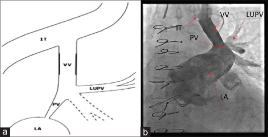
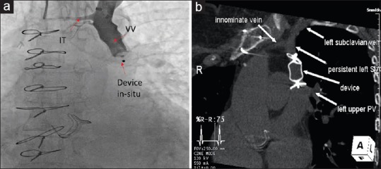
Transcatheter therapy for partial anomalous pulmonary venous connection with dual drainage is unique and rarely reported. We report a 69-year-old female with recurrent brain abscess and partial anomalous connection of the left upper pulmonary vein with dual drainage to the vertical vein (VV) and left atrium (LA). Transcatheter occlusion of the VV was done using an 18-mm St. Jude Amplatzer Vascular Plug II, thus redirecting the left-sided pulmonary venous drainage to LA. Careful evaluation of partial anomalous pulmonary venous drainage with cross-sectional imaging is essential to allow the delineation of dual connections, enabling a less invasive transcatheter treatment approach.
Keywords: Anomalous pulmonary venous connection; device occlusion; dual drainage.
Conflict of interest statement
There are no conflicts of interest.
Figures
References
-
- Ward KE, Mullins CE. Anomalous pulmonary venous connections, vein stenosis, and atresia of the common vein. In: Garson A, Bricker JT, Fisher DJ, Neish SR, editors. The Science and Practice of Pediatric Cardiology. Baltimore, Md: Williams and Wilkins; 1998. pp. 1431–61.
-
- Forbess LW, O’Laughlin MP, Harrison JK. Partially anomalous pulmonary venous connection: Demonstration of dual drainage allowing nonsurgical correction. Cathet Cardiovasc Diagn. 1998;44:330–5. - PubMed
-
- Dähnert I, Riede FT, Kostelka M. Partial anomalous pulmonary venous drainage of the left upper pulmonary vein – Catheter interventional treatment is sometimes possible. Clin Res Cardiol. 2007;96:511–3. - PubMed
-
- Gomez J, Soledispa C. Redirection of anomalous venous pulmonary flow to left atrium using a vascular plug II. J Invasive Cardiol. 2012;24:E96–8. - PubMed
-
- Recto MR, Sadlo H, Sobczyk WL. Rare case of persistent left superior vena cava to left upper pulmonary vein: Pathway for paradoxical embolization and development of transient ischemic attack and subsequent occlusion with an amplatzer vascular plug. J Invasive Cardiol. 2007;19:E313–6. - PubMed