Successful management of a neonate with antenatally detected mature intrapericardial teratoma
- PMID: 31143053
- PMCID: PMC6521665
- DOI: 10.4103/apc.APC_77_18
Successful management of a neonate with antenatally detected mature intrapericardial teratoma
Abstract
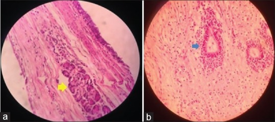
Intrapericardial teratoma is a germ-cell tumor that typically arises from the base of the heart and usually diagnosed in the fetal or neonatal period. Although benign, these tumors can be massive in size causing direct compression of the heart. Life-threatening complications such as fetal hydrops, cardiac failure, superior vena cava syndrome, and cardiac tamponade caused by these teratomas have been reported. Early surgical excision is curative. We present the images of a mature intrapericardial teratoma diagnosed in an asymptomatic neonate. The neonate was managed successfully by elective surgical excision.
Keywords: Germ-cell tumor; intrapericardial teratoma; surgical excision; two-dimensional echocardiography.
Conflict of interest statement
There are no conflicts of interest.
Figures


References
-
- Agozzino L, Vosa C, Arciprete P, de Leva F, Cotrufo M. Intrapericardial teratoma in the newborn. Int J Cardiol. 1984;5:21–8. - PubMed
-
- Manoly I, Viola N, Fowler D, Roman K, Haw M. Intrapericardial teratoma in neonates: A surgical emergency. World J Pediatr Congenit Heart Surg. 2011;2:321–3. - PubMed
-
- Gonzalez-Crussi F. Atlas of Tumor Pathology. Washington: Armed Forces Institute of Pathology; 1982. Extragonadal teratomas; pp. 1–44. 129. 2nd Ser. Fascicle 18.
-
- Sumner TE, Crowe JE, Klein A, McKone RC, Weaver RL. Intrapericardial teratoma in infancy. Pediatr Radiol. 1980;10:51–3. - PubMed
-
- Farooki ZQ, Arciniegas E, Hakimi M, Clapp S, Jackson W, Green EW, et al. Real-time echocardiographic features of intrapericardial teratoma. J Clin Ultrasound. 1982;10:125–8. - PubMed