. 2019 May 9:10:582.
doi: 10.3389/fpls.2019.00582. eCollection 2019.

Anionic Phospholipids Induce Conformational Changes in Phosphoenolpyruvate Carboxylase to Increase Sensitivity to Cathepsin Proteases

Affiliations

Anionic Phospholipids Induce Conformational Changes in Phosphoenolpyruvate Carboxylase to Increase Sensitivity to Cathepsin Proteases

Jacinto Gandullo et al. Front Plant Sci. .

Abstract

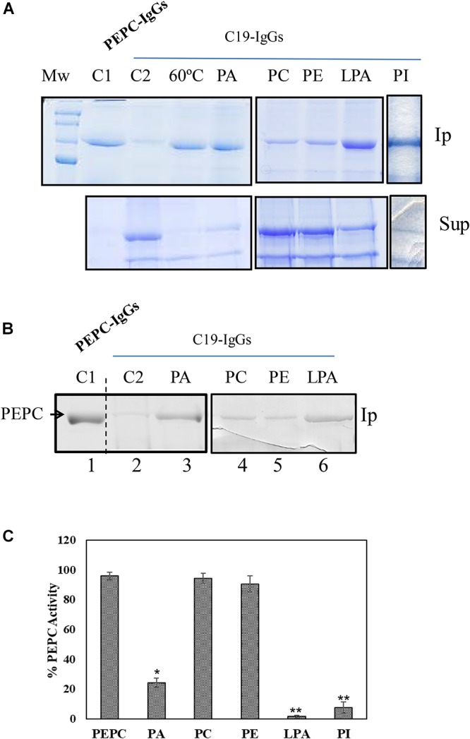
Phosphoenolpyruvate carboxylase (PEPC) is a cytosolic, homotetrameric enzyme that serves a variety of functions in plants, acting as the primary form of CO2 fixation in the C4 photosynthesis pathway (C4-PEPC). In a previous work we have shown that C4-PEPC bind anionic phospholipids, resulting in PEPC inactivation. Also, we showed that PEPC can associate with membranes and to be partially proteolyzed. However, the mechanism controlling this remains unknown. Using semi purified-PEPC from sorghum leaf and a panel of PEPC-specific antibodies, we analyzed the conformational changes in PEPC induced by anionic phospholipids to cause the inactivation of the enzyme. Conformational changes observed involved the exposure of the C-terminus of PEPC from the native, active enzyme conformation. Investigation of the protease activity associated with PEPC demonstrated that cysteine proteases co-purify with the enzyme, with protease-specific substrates revealing cathepsin B and L as the major protease species present. The anionic phospholipid-induced C-terminal exposed conformation of PEPC appeared highly sensitive to the identified cathepsin protease activity and showed initial proteolysis of the enzyme beginning at the N-terminus. Taken together, these data provide the first evidence that anionic phospholipids promote not only the inactivation of the PEPC enzyme, but also its proteolysis.

Keywords: cathepsin proteases; conformational changes; phosphatidic acid; phosphoenolpyruvate carboxylase; phospholipids; proteolysis; sorghum.


Figures

Figure 1
Figure 1
Anionic phospholipids promoted extensive conformational changes in PEPC detected by the exposure of its C-terminus. 0.2 U (A) or 0.1 U (B) of PEPC were incubated in the absence (lanes C1, C2 and 60°C) or presence of 0.25 mM of the phospholipids indicated (lanes PA, PC, PE, LPA, PI). Following 30 min incubation at 30°C, PEPC was immunoprecipitated with PEPC-IgG (lane C1) or C19-IgG (remaining samples). An aliquot of PEPC was heated at 60°C for 2 min to serve as a positive control for C19-IgG (lane 60°C). This treatment completely exposes the C-terminus of the protein (Alvarez et al., 2003). (A) Immunoprecipitates (Ip) and supernatant (Sup) were analyzed by SDS–PAGE (10%) and stained with Coomassie blue. (B) Immunoprecipitates were immunoblotted with PEPC-IgG. The images in (A) and (B) are representative of at least 3 independent experiments. (C) Effect of the different phospholipids on PEPC activity. PA, phosphatidic acid; PC, phosphatidylcholine; PE, phosphatidylethanolamine; LPA, lyso-PA; PI, phosphatidylinositol4-P. Data represent mean ± SE of 3–6 different experiments. Statistically significant difference with respect controls are indicated by () at P < 0.05, (∗∗) at P < 0.01, using the Dunnett test.
Figure 2
Figure 2
Effect of pH, enzyme phosphorylation state and Glc-6P on the exposure of the C-terminus of C4-PEPC. (A) Phosphorylated (OP) or non-phosphorylated (OH) sp-PEPC were incubated in 0.1 M Tris–HCl buffer at different pHs (8, 7,3 or 7,1). Following 30 min at 30°C, PEPC was immunoprecipitated with C19-IgG. Lane C, sp-PEPC prior to immunoprecipitation. Phosphorylated PEPC was obtained by in vitro phosphorylation with PKA as described in M&M. (B) Non-phosphorylated PEPC incubated with 5 mM Glc-6P for 30 min and then immunoprecipitated with C19-IgG. (C) Clarified protein extract from illuminated (L, phosphorylated-PEPC) or dark (D, non-phosphorylated-PEPC) sorghum leaves incubated in the presence of Glc-6P or L-malate and immunoprecipitated with PEPC-IgG (lanes 1, 2) or C19-IgG (lanes 3–7). Protein was detected on immunoblots using PEPC-IgG. Ip, immunoprecipitated; Sup, supernatant. The enzyme phosphorylation state was controlled via the L-malate sensitivity test (Table 1).
Figure 3
Figure 3
SDS–PAGE analysis of the sp-PEPC fraction during the purification process. PEPC (0.2 U) from the different purification steps were subjected to SDS–PAGE and staining with Commassie blue. Lane 1, clarified protein extract from dark-adapted sorghum leaves; Lanes 2, 3, chromatografhy on Hydroxyapatite and on anion-exchange, respectively. Lane 3 correspond to the final sp-PEPC preparation mainly used in the experiments. Lanes 1–3 contain 0.2 U of PEPC). Lane 4, immunoblot of lane 3 (0.05 U PEPC) detected using PEPC-IgG. Lane 5, immunoblot of sp-PEPC (0.1 U PEPC) following 3 months at –20°C.
Figure 4
Figure 4
Anionic phospholipids promote proteolysis of C4-PEPC. (A) Sp-PEPC was incubated in the absence (lane C) or in the presence of 0.25 mM of the different phospholipids (lanes PA, PI, PC, PE, LPA) at 30°C as is described in M&M. (B) Range of PA concentration promoting PEPC proteolysis. A standard proteolytic assay was performed with increasing concentrations of PA18:1. At the indicated times, 0.05 U PEPC aliquots were removed and subjected to immunoblot. Bands were detected with PEPC-IgG.
Figure 5
Figure 5
The N-terminal of PEPC is the initial target of PA-induced proteolysis. A standard proteolytic assay as is described in M&M was performed in presence or absence of 0.25 mM PA. (A) Coomassie blue stained PEPC samples exposed to PA for 60 min at 30°C and immunoprecipitated with C19-IgG. Lane 1, PEPC prior to immunoprecipitation. Lane 2, PEPC without PA. Lane 3, PEPC in presence of PA; (B) Immunoblot analysis of PEPC exposed to PA at different times and detected using N24-IgG or PEPC-IgG. Lane 1, PEPC control without PA. Lanes 2 and 3 in B, 5 or 7 h of incubation in presence of PA, respetively.
Figure 6
Figure 6
(A) sp-PEPC exposed to PA in the presence of protease inhibitors: sp-PEPC control (lane 1), no protease inhibitors (lane 2), 1 mM Bestatin (lane 3), 0.2 mM Leupeptin (lane 4), 0.065 mM Chymostatin (lane 5), 1 mM PMSF (lane 6), Sigma protease inhibitor cocktail containing AEBSF, 1,10-Phenanthroline, Pepstatin, Leupeptin, Bestatin and E-64 (lane 7) or 0.1 mM Aprotinin (lane 8). Following 20 h of incubation at 30°C, samples were subjected to SDS–PAGE and stained with Commassie Blue. (B) Cistatin and E-64 inhibit the protease activity present in sp-PEPC. Aliquots of sp-PEPC (25 μl) were incubated with 1 μM of recombinant purified cystatin (CIP) or E-64 in a 0.1 M citrate buffer (pH 6), 0.15 M NaCl and 5 mM MgCl2 for 10 min at 30°C. Protease activity was assayed by addition of 2 mM of the cathepsin L-specific substrate, Z-FR-AMC. The reaction was developed 5 h at 30°C. Protease activity is expressed as % with respect to the control in absence of inhibitor (sp-PEPC). Date are the mean ± SE of triplicate measurement and statistically significant difference with respect to the control are indicated by () at P < 0.05, (∗∗) at P < 0.01, using the Dunnett test.

References

    1. Alvarez R., García-Mauriño S., Feria A.-B., Vidal J., Echevarría C. (2003). A conserved 19-amino acid synthetic peptide from the carboxy terminus of phosphoenolpyruvate carboxylase inhibits the in vitro phosphorylation of the enzyme by the calcium-independent phosphoenolpyruvate carboxylase kinase. Plant Physiol. 132 1097–1106. 10.1104/pp.103.023937 - DOI - PMC - PubMed
    1. Baur B., Dietz K. J., Winter K. (1992). Regulatory protein phosphorylation of phosphoenolpyruvate carboxylase in the facultative crassulacean-acid-metabolism plant: Mesembryanthemum crystallinum L. Eur. J. Biochem. 209 95–101. 10.1111/j.1432-1033.1992.tb17265.x - DOI - PubMed
    1. Bradford M. M., Dong Y., Xu L., Liu S., Bai X., Bradford M., et al. (1976). A rapid and sensitive method for the quantitation of microgram quantities of protein utilizing the principle of protein-dye binding. Anal. Biochem. 72 248–254. 10.1016/0003-2697(76)90527-90523 - DOI - PubMed
    1. Carrillo L., Martínez M., Ramessar K., Cambra I., Castañera P., Ortego F., et al. (2011). Expression of a barley cystatin gene in maize enhances resistance against phytophagous mites by altering their cysteine-proteases. Plant Cell Rep. 30 101–112. 10.1007/s00299-010-0948-z - DOI - PubMed
    1. Chollet R., Vidal J., O’Leary M. H. (1996). Phosphoenolpyruvate carboxylase: a ubiquitous, highly regulated enzyme in plants. Annu. Rev. Plant Physiol. Plant Mol. Biol. 47 273–298. 10.1146/annurev.arplant.47.1.273 - DOI - PubMed