White Epidermoid of the Sylvian Fissure Masquerading as a Dermoid Cyst: An Extremely Rare Occurrence
- PMID: 31143281
- PMCID: PMC6515996
- DOI: 10.4103/ajns.AJNS_241_18
White Epidermoid of the Sylvian Fissure Masquerading as a Dermoid Cyst: An Extremely Rare Occurrence
Abstract
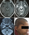
We report the case of a 30-year-old female with a Sylvian fissure, white epidermoid which was radiologically looking like a dermoid cyst. The female presented with a headache with no neurological deficits. On radiology, the lesion was in Sylvian fissure, T1 hyperintense, T2 hypointense, and with minimal diffusion restriction medially. Hence a preoperative impression of dermoid cyst was made, a quite uncommon location. Intraoperatively, the classical pearly-white flaky appearance of epidermoid was seen which was confirmed histopathologically. White epidermoids appearing so because of high protein content are a rarity and are more likely to cause aseptic meningitis in the event of intraoperative spillage. Differentiating between a dermoid cyst and white epidermoid preoperatively and radiologically is difficult. Dermoids show diffusion restriction and are usually midline, whereas white epidermoids do not show diffusion restriction and are usually lateral. This is the first report of a white epidermoid in Sylvian fissure to the best of our knowledge.
Keywords: Dermoid cyst; Sylvian fissure; mimicking; white epidermoid.
Conflict of interest statement
There are no conflicts of interest.
Figures


References
-
- Puranik A, Sankhe S, Goel N, Mahore A. Cerebral shading sign in a giant intraparenchymal white epidermoid. Neurol India. 2012;60:265–6. - PubMed
-
- Conley FK. Epidermoid and dermoid tumors: Clinical features and surgical management. In: Wilkins RH, Rengachary SS, editors. Neurosurgery. 2nd ed. United States of America: McGraw-Hill; 1996. p. 971.
-
- Goel A, Muzumdar D, Desai K. Anterior tentorium-based epidermoid tumours: Results of radical surgical treatment in 96 cases. Br J Neurosurg. 2006;20:139–45. - PubMed
-
- Garces J, Mathkour M, Beard B, Sulaiman OA, Ware ML. Insular and sylvian fissure dermoid cyst with giant cell reactivity: Case report and review of literature. World Neurosurg. 2016;93:491.e1–5. - PubMed

