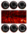
Multimodal endoscopy for colorectal cancer detection by optical coherence tomography and near-infrared fluorescence imaging
- PMID: 31143497
- PMCID: PMC6524571
- DOI: 10.1364/BOE.10.002419
Multimodal endoscopy for colorectal cancer detection by optical coherence tomography and near-infrared fluorescence imaging
Abstract
While colonoscopy is the gold standard for diagnosis and classification of colorectal cancer (CRC), its sensitivity and specificity are operator-dependent and are especially poor for small and flat lesions. Contemporary imaging modalities, such as optical coherence tomography (OCT) and near-infrared (NIR) fluorescence, have been investigated to visualize microvasculature and morphological changes for detecting early stage CRC in the gastrointestinal (GI) tract. In our study, we developed a multimodal endoscopic system with simultaneous co-registered OCT and NIR fluorescence imaging. By introducing a contrast agent into the vascular network, NIR fluorescence is able to highlight the cancer-suspected area based on significant change of tumor vascular density and morphology caused by angiogenesis. With the addition of co-registered OCT images to reveal subsurface tissue layer architecture, the suspected regions can be further investigated by the altered light scattering resulting from the morphological abnormality. Using this multimodal imaging system, an in vivo animal study was performed using a F344-ApcPircUwm rat, in which the layered architecture and microvasculature of the colorectal wall at different time points were demonstrated. The co-registered OCT and NIR fluorescence images allowed the identification and differentiation of normal colon, hyperplastic polyp, adenomatous polyp, and adenocarcinoma. This multimodal imaging strategy using a single imaging probe has demonstrated the enhanced capability of identification and classification of CRC compared to using any of these technologies alone, thus has the potential to provide a new clinical tool to advance gastroenterology practice.
Conflict of interest statement
Dr. Chen has a financial interest in OCT Medical Imaging, Inc., which, however, did not support this work.
Figures


Similar articles
-
Multimodality endoscopic optical coherence tomography and fluorescence imaging technology for visualization of layered architecture and subsurface microvasculature.Opt Lett. 2018 May 1;43(9):2074-2077. doi: 10.1364/OL.43.002074. Opt Lett. 2018. PMID: 29714749 Free PMC article.
-
Molecular Fluorescence Endoscopy Targeting Vascular Endothelial Growth Factor A for Improved Colorectal Polyp Detection.J Nucl Med. 2016 Mar;57(3):480-5. doi: 10.2967/jnumed.115.166975. Epub 2015 Dec 17. J Nucl Med. 2016. PMID: 26678613
-
In vivo, dual-modality OCT/LIF imaging using a novel VEGF receptor-targeted NIR fluorescent probe in the AOM-treated mouse model.Mol Imaging Biol. 2011 Dec;13(6):1173-82. doi: 10.1007/s11307-010-0450-6. Mol Imaging Biol. 2011. PMID: 21042865 Free PMC article.
-
Optical coherence tomography in detection of dysplasia and cancer of the gastrointestinal tract and bilio-pancreatic ductal system.World J Gastroenterol. 2008 Nov 14;14(42):6444-52. doi: 10.3748/wjg.14.6444. World J Gastroenterol. 2008. PMID: 19030194 Free PMC article. Review.
-
Optical coherence tomography.ScientificWorldJournal. 2007 Jan 26;7:87-108. doi: 10.1100/tsw.2007.29. ScientificWorldJournal. 2007. PMID: 17334603 Free PMC article. Review.
Cited by
-
Diagnosing colorectal abnormalities using scattering coefficient maps acquired from optical coherence tomography.J Biophotonics. 2021 Jan;14(1):e202000276. doi: 10.1002/jbio.202000276. Epub 2020 Oct 22. J Biophotonics. 2021. PMID: 33064368 Free PMC article.
-
Advances in Endoscopic Photoacoustic Imaging.Photonics. 2021 Jul;8(7):281. doi: 10.3390/photonics8070281. Epub 2021 Jul 16. Photonics. 2021. PMID: 35252433 Free PMC article.
-
Biomedical optical imaging technology and applications: From basic research toward clinical diagnosis.Exp Biol Med (Maywood). 2020 Feb;245(4):269-272. doi: 10.1177/1535370220909543. Exp Biol Med (Maywood). 2020. PMID: 32141780 Free PMC article. No abstract available.
-
Targeted Biodegradable Near-Infrared Fluorescent Nanoparticles for Colorectal Cancer Imaging.ACS Appl Bio Mater. 2024 Dec 16;7(12):7861-7870. doi: 10.1021/acsabm.4c00072. Epub 2024 Apr 4. ACS Appl Bio Mater. 2024. PMID: 38574012 Free PMC article.
-
In vivo functional imaging of the human middle ear with a hand-held optical coherence tomography device.Biomed Opt Express. 2021 Jul 22;12(8):5196-5213. doi: 10.1364/BOE.430935. eCollection 2021 Aug 1. Biomed Opt Express. 2021. PMID: 34513251 Free PMC article.
References
-
- Stewart B. W., Wild C., International Agency for Research on Cancer, and World Health Organization, World cancer report 2014, pp. xiv, 630 pages.
Grants and funding
LinkOut - more resources
Full Text Sources
Miscellaneous
